Download
1 / 35
350 likes | 667 Vues
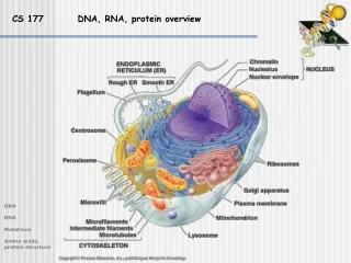
CS 177 DNA, RNA, protein overview. DNA RNA Mutations Amino acids, protein structure. DNA, RNA, protein overview. Questions about the genome in an organism: How much DNA, how many nucleotides? How many genes are there? What types of proteins appear to be coded by these genes?.

E N D
CS 177 DNA, RNA, protein overview DNA RNA Mutations Amino acids, protein structure
DNA, RNA, protein overview Questions about the genome in an organism: How much DNA, how many nucleotides? How many genes are there? What types of proteins appear to be coded by these genes? Questions about the proteome: What proteins are present? Where are they? When are they present - under what conditions? DNA RNA Mutations Amino acids, protein structure
DNA, RNA, protein overview Lecture 2 * DNA and its components * RNA and its components * Mutations * Amino acids, review of protein structure DNA RNA Mutations Amino acids, protein structure
Adenine Thymine Cytosine Guanine DNA overview DNA deoxyribonucleic acid 4 bases Pyrimidine (C4N2H4) Pyrimidine (C4N2H4) Purine (C5N4H4) A = T = C = G = Thymine Cytosine Nucleoside Nucleotide base + sugar (deoxyribose) base + sugar + phosphate DNA RNA Mutations Amino acids, protein structure Numbering of carbons?
Linking nucleotides Hydrogen bonds N-H------N N-H------O Linking nucleotides: The 3’-OH of one nucleotide is linked to the 5’-phosphate of the next nucleotide What next? Thymine Adenine Cytosine DNA RNA Mutations Amino acids, protein structure Guanine
Base pairing A T Base pairing (Watson-Crick): A/T (2 hydrogen bonds) G/C (3 hydrogen bonds) C G Always pairing a purine and a pyrimidine yields a constant width A T DNA base composition: A + G = T + C (Chargaff’s rule) T A DNA RNA Mutations Amino acids, protein structure C G
DNA conventions 1. DNA is a right-handed helix DNA RNA Mutations Amino acids, protein structure
DNA conventions 1. DNA is a right-handed helix 2. The 5’ end is to the left by convention 5’ 3’ -ATCGCAATCAGCTAGGTT- sense (forward) 3’ 5’ -TAGCGTTAGTCGATCCAA- antisense (reverse) 5’-ATCGCAATCAGCTAGGTT-3’ 3’-TAGCGTTAGTCGATCCAA-5’ DNA RNA Mutations Amino acids, protein structure
DNA structure Some more facts: 1. Forces stabilizing DNA structure: Watson-Crick-H-bonding and base stacking(planar aromatic bases overlap geometrically and electronically energy gain) 2. Genomic DNAs are large molecules:Eschericia coli: 4.7 x 10⁶ bp; ~ 1 mm contour length Human: 3.2 x 10⁹ bp; ~ 1 m contour length 3. Some DNA molecules (plasmids) are circular and have no free ends: mtDNA bacterial DNA (only one circular chromosome) 4. Average gene of 1000 bp can code for average protein of about 330 amino acids 5. Percentage of non-coding DNA varies greatly among organisms Organism # Base pairs # Genes Non-coding DNA small virus 4 x 10³ 3 very little ‘typical’ virus 3 x 10⁵ 200 very little DNA RNA Mutations Amino acids, protein structure bacterium 5 x 10⁶ 3000 10 - 20% yeast 1 x 10⁷ 6000 > 50% human 3.2 x 10⁹ 30,000? 99% amphibians < 80 x 109 ? ? plants < 900 x 109 23,000 - >50,000 > 99%
RNA structure RNA Pyrimidine (C4N2H4) Purine (C5N4H4) Adenine Uracil Cytosine Guanine Thymine (DNA) Uracil (RNA) Nucleoside Nucleotide base + sugar (ribose) base + sugar + phosphate 3 major types of RNA messenger RNA (mRNA); template for protein synthesis transfer RNA (tRNA); adaptor molecules that decode the genetic coderibosomal RNA (rRNA); catalyzing the synthesis of proteins ribonucleic acid 4 bases A = U = C = G = DNA RNA Mutations Amino acids, protein structure
Base interactions in RNA Base pairing: U/A/(T) (2 hydrogen bonds) G/C (3 hydrogen bonds) RNA base composition: A + G = U + C / Chargaff’s rule does not apply (RNA usually prevails as single strand) RNA structure: - usually single stranded - many self-complementary regions RNA commonly exhibits an intricate secondary structure (relatively short, double helical segments alternated with single stranded regions) - complex tertiary interactions fold the RNA in its final three dimensional form - the folded RNA molecule is stabilized by interactions (e.g. hydrogen bonds and base stacking) DNA RNA Mutations Amino acids, protein structure
A) single stranded regions B) duplex C C) hairpin D) internal loop D E) bulge loop G E F F) junction B A G) pseudoknot RNA structure Primary structure formed by unpaired nucleotides Secondary structure double helical RNA (A-form with 11 bp per turn) duplex bridged by a loop of unpaired nucleotides nucleotides not forming Watson-Crick base pairs unpaired nucleotides in one strand,other strand has contiguous base pairing DNA RNA Mutations Amino acids, protein structure three or more duplexes separated by singlestranded regions tertiary interaction between bases of hairpin loopand outside bases
RNA structure Primary structure Secondary structure Tertiary structure C D G E F DNA RNA Mutations Amino acids, protein structure B A
RNA structure How to predict RNA secondary/tertiary structure? Probing RNA structure experimentally: - physical methods (single crystal X-ray diffraction, electron microscopy) - chemical and enzymatic methods - mutational analysis (introduction of specific mutations to test change in some function or protein-RNA interaction) Thermodynamic prediction of RNA structure: - RNA molecules comply to the laws of thermodynamics, therefore it should be possible to deduce RNA structure from its sequence by finding the conformation with the lowest free energy - Pros: only one sequence required; no difficult experiments; does not rely on alignments - Cons: thermodynamic data experimentally determined, but not always accurate; possible interactions of RNA with solvent, ions, and proteins Comparative determination of RNA structure: - basic assumption: secondary structure of a functional RNA will be conserved in the evolution of the molecule (at least more conserved than the primary structure); when a set of homologous sequences has a certain structure in common, this structure can be deduced by comparing the structures possible from their sequences - Pros: very powerful in finding secondary structure, relatively easy to use, only sequences required, not affected by interactions of the RNA and other molecules - Cons: large number of sequences to study preferred, structure constrains in fully conserved regions cannot be inferred, extremely variable regions cause problems with alignment DNA RNA Mutations Amino acids, protein structure
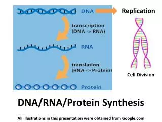
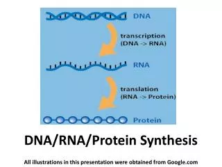
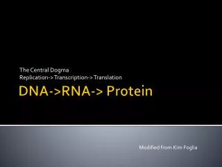
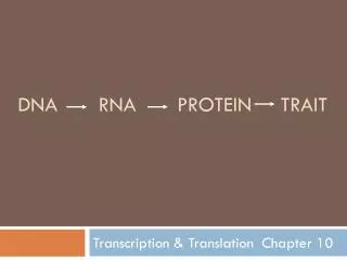
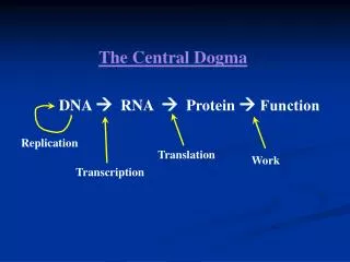
Amino acids/proteins The “central dogma” of modern biology: DNA RNA protein Getting from DNA to protein: Two parts: 1. Transcription in which a short portion of chromosomal DNA is used to make a RNA molecule small enough to leave the nucleus. 2. Translation in which the RNA code is used to assemble the protein at the ribosome The genetic code - The code problem: 4 nucleotides in RNA, but 20 amino acids in proteins - Bases are read in groups of 3 (= a codon) - The code consists of codons 64 (43 = 64) - All codons are used in protein synthesis: - 20 amino acids - 3 stop codons - AUG (methionine) is the start codon (also used internally) - The code is non-overlapping and punctuation-free DNA RNA Mutations Amino acids, protein structure - The code is degenerate (but NOT ambiguous): each amino acid is specified by at least one codon - The code is universal (virtually all organisms use the same code)
In-class exercise 1. Which amino acids are specified by single codons? 2. How many amino acids are specified by the first two nucleotides only? 3. What is the RNA code for the start codon? The genetic code methionine and tryptophan five: proline, threonine,valine, alanine, glycine DNA RNA Mutations Amino acids, protein structure AUG
Amino acids Hydrophobic G A V L I DNA RNA Mutations Amino acids, protein structure M F W P
Amino acids Hydrophyllic S T C Y N Q DNA RNA Mutations Amino acids, protein structure D E K R H
- a reading frame is not always easily recognizable - each strand of RNA/DNA has three possible starting points (position one, two, or three): Position 1 CAG AUG AGG UCA GGC AUA gln met arg ser gly ile Position 2 C AGA UGA GGU CAG GCA UA arg trp gly gln ala Position 3 CA GAU GAG GUC AGG CAU A asp glu val arg his Up to 30% of mutations causing humane disease are due to premature termination of translation (nonsense mutations or frameshift) Reading frames Reading frame (also open reading frame): The stretch of triplet sequence of DNA that potentially encodes a protein. The reading frame is designated by the initiation or start codon and is terminated by a stop codon. - mutations within an open reading frame that delete or add nucleotides can disrupt the reading frame (frameshift mutation): DNA RNA Mutations Amino acids, protein structure Wildtype CAG AUG AGG UCA GGC AUA GAG gln met arg ser gly ile glu Mutant CAG AUG AGU CAG GCA UAG AG gln met ser gln ala
Mutations Mutation: any heritable change in DNA Sources of mutation: Spontaneous mutations: mutations occur for unknown reasons Induced mutations: exposure to substance (mutagen) known to cause mutations, e.g. X-rays, UV light, free radicals Mutations may influence one or several base pairs a) Nucleotide substitutions (point mutation) 1) Transitions (Pu Pu; Py Py)2) Transversions (Pu Py) In-class exercise How many transition and transversion events are possible? 2 transitions: T C; A G4 transversions: T A; T G C A; C G b) Insertion or deletion (“indels”) - one to many bases can be involved - frequently associated with repeated sequences (“hot spots”) - lead to frameshift in protein-coding genes, except when N = 3X - also caused by insertion of transposable elements into genes DNA RNA Mutations Amino acids, protein structure “Weighting” of mutation events plays important role for phylogenetic analyses (model of sequence evolution)
Mutations Mutations may influence phenotype a) Silent (or synonymous) substitution - nucleotide substitution without amino acid change- no effect on phenotype- mostly third codon position- other possible silent substitutions: changes in non-coding DNA b) Replacement substitution - causes amino acid change - neutral: protein still functions normally - missense: protein loses some functions (e.g. sickle cell anemia: mutation in ß-globin) c) Sense/nonsense substitution - sense: involves a change from a termination codon to one that codes for an amino acid - nonsense: creates premature termination codon Mutation rates = a measure of the frequency of a given mutation per generation - mutation rates are usually given for specific loci (e.g. sickle cell anemia)- the rate of nucleotide substitutions in humans is on the order of 1 per 100,000,000- range varies from 1 in 10,000 to 1 in 10,000,000,000- every human has about 30 new mutations involving nucleotide substitutions- mutation rate is about twice as high in male as in female meiosis DNA RNA Mutations Amino acids, protein structure
Mutations A single amino acid substitution in a protein causes sickle-cell disease DNA RNA Mutations Amino acids, protein structure
Review of protein structure DNA RNA Mutations Amino acids, protein structure Making a polypeptide chain
Polypeptide chain Review of protein structure Primary structure Proteins are chains of amino acids joined by peptide bonds The structure of two amid acids The N-C-C sequence is repeated throughout the protein, forming the backbone The bonds on each side of the C atom are free to rotate within spatial constrains,the angles of these bonds determine the conformation of the protein backbone DNA RNA Mutations Amino acids, protein structure The R side chains also play an important structural role
Review of protein structure Secondary structure: Interactions that occur between the C=O and N-H groups on amino acidsMuch of the protein core comprises helices and sheets, folded into a three-dimensional configuration:- regular patterns of H bonds are formed between neighboring amino acids- the amino acids have similar angles- the formation of these structures neutralizes the polar groups on each amino acid- the secondary structures are tightly packed in a hydrophobic environment- Each R side group has a limited volume to occupy and a limited number of interactions with other R side groups sheet helix DNA RNA Mutations Amino acids, protein structure
Secondary structure Other Secondary structure elements(no standardized classification) - random coil - loop - others (e.g. 310 helix, -hairpin, paperclip) Super-secondary structure - In addition to secondary structure elements that apply to all proteins (e.g. helix, sheet) there are some simple structural motifs in some proteins DNA RNA Mutations Amino acids, protein structure - These super-secondary structures (e.g. transmembrane domains, coiled coils, helix-turn-helix, signal peptides) can give important hints about protein function
Secondary structure Structural classification of proteins (SCOP) Class 2: mainly beta Class 1: mainly alpha Class 3: alpha/beta Class 4: few secondary structures DNA RNA Mutations Amino acids, protein structure
Secondary structure Alternative SCOP Class : antiparallel sheets Class / : mainly sheetswith intervening helices Class : only helices Class + : mainlysegregated helices withantiparallel sheets Membrane structure:hydrophobic helices withmembrane bilayers Multidomain: containmore than one class DNA RNA Mutations Amino acids, protein structure
A: No, because the detailed packing of the amino acid side chains is not revealed from this information. However, the Psi and Phi angles do determine the entire secondary structure of a protein Tertiary structure Review of protein structure Q: If we have all the Psi and Phi angles in a protein, do we then have enough information to describe the 3-D structure? DNA RNA Mutations Amino acids, protein structure
Tertiary structure The tertiary structure describes the organization in three dimensionsof all the atoms in the polypeptide The tertiary structure is determined by a combination of different types of bonding(covalent bonds, ionic bonds, h-bonding, hydrophobic interactions, Van der Waal’s forces) between the side chains Many of these bonds are very week and easy to break, but hundreds or thousands working together give the protein structure great stability If a protein consists of only one polypeptide chain, this level then describes thecomplete structure DNA RNA Mutations Amino acids, protein structure
Tertiary structure Proteins can be divided into two general classes based on their tertiary structure: - Fibrous proteins have elongated structure with the polypeptide chains arranged in long strands. This class of proteins serves as major structural component of cells Examples: silk, keratin, collagen - Globular proteins have more compact, often irregular structures. This class of proteins includes most enzymes and most proteins involved in gene expression and regulation DNA RNA Mutations Amino acids, protein structure
Quaternary structure The quaternarystructure defines the conformation assumed by a multimeric protein.The individual polypeptide chains that make up a multimeric protein are often referred toas protein subunits. Subunits are joined by ionic, H and hydrophobic interactions Example:Haemoglobin(4 subunits) DNA RNA Mutations Amino acids, protein structure
Structure displays Common displays are (among others) cartoon, spacefill, and backbone backbone spacefill cartoon DNA RNA Mutations Amino acids, protein structure
Primary structure: Sequence of amino acids Secondary structure: Interactions that occur betweenthe C=O and N-H groups on amino acids Tertiary structure: Organization in three dimensions of all the atoms in the polypeptide Quaternary structure: Conformation assumed by a multimeric protein The four levels of protein structure are hierarchical:each level of the build process is dependent upon the one below it Summary protein structure DNA RNA Mutations Amino acids, protein structure
Next week First quiz Lecture 1 - Bioinformatics definitions - The human genome project Lecture 2 • - DNA structure • - RNA structure • - Mutations • Amino acids • - Proteins DNA RNA Mutations Amino acids, protein structure