The Reproductive Systems
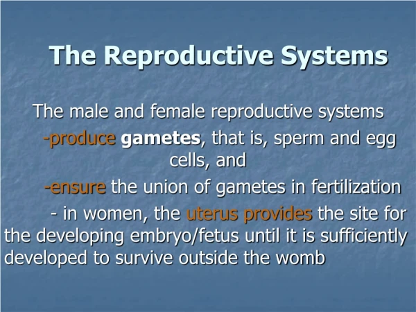
The Reproductive Systems. Sexual Reproduction. Offspring produced by the joining (fertilization) of gametes Requires halving of chromosomes (46 23) through meiosis Diploid haploid Separation of homologous chromosomes Autosomes vs. sex chromosomes

The Reproductive Systems
E N D
Presentation Transcript
Sexual Reproduction • Offspring produced by the joining (fertilization) of gametes • Requires halving of chromosomes (4623) through meiosis • Diploid haploid • Separation of homologous chromosomes • Autosomes vs. sex chromosomes • Variability provided by fertilization, independent assortment of homologs, crossing-over (genetic recombination)
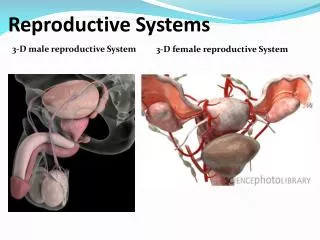
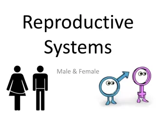
Reproductive Structures • Gonads - gamete production • Ducts - storage, maturation and transport of gametes • Accessory sex glands - secretions that maintain gametes or aid in copulation • Supporting structures - structures important in copulation or fetal development
Testicular Structure • Contained in scrotum divided in two by fascia and dartos muscle (smooth) • Tunica vaginalis - derived from peritoneum during decent through inguinal canal • Tunica albuginea - protrudes interiorly dividing testes in lobular arrangement
More Testicular Structure • Lobules contain seminiferous tubules - wall lined with spermatogenic cells (any stage) and sustentacular (Sertoli) cells • Lobules also contain interstitial endocrinocytes (Leydig cells - secrete testosterone)
Spermatogenic Cells • Spermatogonia arranged at basement membrane of tubule, mitotically divide to form more spermatogonia (2n, source of endless supply) • Some lose contact with basement membrane and differentiate to primary spermatocytes (2n) • Continuing “maturity” with distance from basement membrane
More Spermatogenic Cells • Secondary spermatocyte - result of first meiotic division (n but with chromatid duplicates) • Spermatid - result of second meiotic division (n) • Meiotic divisions don’t complete cytokinesis - cytoplasmic bridges between cells • Sperm or spermatozoa - result of spermiogenesis • Formation of acrosome and flagellum
Spermatozoa • 300 million produced per day • Head - DNA and acrosome (hyaluronidase and proteinases) • Midpiece - mitochondria for ATP production • Tail - flagellum
Sustentacular Cells • Large cells that reach from basement membrane to tubular lumen • Tight junctions between adjacent cells causing blood-testis barrier • Important since sperm have foreign surface antigens which could cause immune response
More Sustentacular Cells • Variety of functions - control and protect spermatogenic cells, nourish developing sperm, phagocytize excess cytoplasm of developing sperm, influence effects of testosterone and FSH, produce fluid for sperm transport, secrete inhibin which regulates sperm production through negative feedback to FSH secretion
Hormonal Control • GnRH produced by hypothalamus - increased at puberty • Stimulates anterior pituitary production of LH and FSH • LH stimulates interstitial endocrinocyte production of testosterone • Lipid soluble - derived from cholesterol • Some target cells (e.g. prostate) covert it to dihydrotestosterone (DHT) - more potent form • Both alter gene action
More Hormonal Control • FSH stimulates spermatogenesis in concert with testosterone • Stimulates sustentacular cells to secrete androgen-binding protein (ABP) which hold testosterone in midst of spermatogenic cells (aiding in the completion of spermatogenesis) • Blood testosterone levels control GnRH • Inhibin from sustentacular cells control FSH
Hormonal Effects • Fetal development • Testosterone - internal structures and testicular descent • DHT - external genitals • Sexual characteristics at puberty • Enlargement of genitals, skeletal and muscle growth, body hair, increased sebaceous secretion, enlargement of larynx
More Hormonal Effects • Sexual function - spermatogenesis and sexual behavior • Metabolic function - protein anabolism (synthesis)
Male Ducts • Seminiferous tubules straight tubules rete testis efferent ducts ductus epididymis ductus deferens ejaculatory duct urethra • Ductus epididymis - site of maturation (increased motility), storage (month), reabsorption of aging sperm, and peristaltic push into ductus deferens • Ductus deferens (distal expansion - ampulla) - storage (several months) and peristaltic push (three smooth muscle layers)
More Male Ducts • Spermatic cord passes through inguinal canal - hernia • Vasectomy • Ejaculatory duct - combination of sperm and fluids from seminal vesicle, eject sperm into urethra • Prostatic, membranous, and spongy urethra • Ejaculation - sympathetic reflex - involves peristaltic contraction from ampulla through spongy urethra
Male Accessory Sex Glands • Seminal vesicle - alkaline fluid with fructose (for ATP), prostaglandins (motility) and clotting proteins (unknown) • 60% of semen • Prostate gland - milky, acidic with citrate (for ATP), acid phosphatase (unknown) and proteolytic enzymes including prostate-specific antigen(PSA) (liquefy clotted sperm) • 25% of semen
More Accessory Glands • Enlarges after age 45 • Prostate cancer • Bulbourethral (Cowper’s) gland - alkaline secretion during arousal and lubricating mucus
Semen Analysis • Fertility evaluation • Volume - 2.5-5 ml • Count - 20 million/ml (typically between 50-150) • Motility - 60% • Morphology - <35% abnormality • pH - 7.2 to 7.7 • Fructose - presence
Male Supporting Structures • Scrotum • Testicular sac composed of loose skin, superficial fascia and dartos muscle (smooth), divided in two by septum • Cremaster muscle (skeletal) - continuation of internal oblique, passing through spermatic cord • Contraction of muscles important to regulation of scrotal temperature (3°C below body temp)
More Male Supporting Structures • Penis • Erectile tissue - corpora cavernosa, corpus spongiosum with tunica albuginea • Erection is a parasympathetic reflex causing vasodilation of arterioles • Glans penis, prepuce (foreskin) • Bulb and crura of penis - points of muscular attachment which aid ejaculation
Ovarian Structure • Held in place by ligaments to uterus and pelvic cavity wall • Germinal epithelium - surrounds ovary & continuous with layer of broad ligament • Tunica albuginea - dense, irregular tissue capsule • Stroma - connective tissue structure, cortex is dense, medulla more loose • Ovarian follicles - in varying stages • Corpus luteum - degenerating follicle
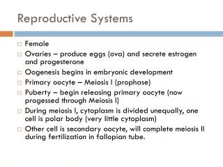
Oogenesis • Primordial germ cells migrate and differentiate oogonia (2n) during development • Primary oocytes (in prophase of meiosis I) • Surrounded by layer of follicular cells inclusively called primordial follicle • 200K to 2M of oogonia and primary oocytes in each ovary at birth • Primary follicles - additional layers of granulosa cells with separating zona pellucida layer • Production begins during childhood (pre-puberty)
More Oogenesis • Outer granulosa cells against basement membrane forming theca folliculi • Theca folliculi further develops into theca interna and externa • Secondary follicles - granulosa cells secrete follicular fluid forming antrum • These present at puberty • Other oocytes degenerate - atresia • Secondary oocyte (n but with chromatid duplicates) - result of completion of meisois I, second cell is smaller (first polar body)
Even More Oogenesis • Graafian (mature) follicle - result of advance to metaphase of meiosis II • At ovulation, secondary oocyte released (with 1st polar body), if fertilized, completes meiosis II (n) and 2 more polar bodies formed
Uterine Tubes • Or oviducts or Fallopian tubes • Carry secondary oocytes to uterus, also frequent site of fertilization • Infundibulum with fimbriae on perimeter • Three layers • Mucosa - ciliated columnar epithelium and secretory cells • Muscularis - peristaltic smooth muscle contraction • Serosa • Tubal ligation
Uterus • Site of fertilized egg implantation and fetal development, source of menstrual flow • Fundus, body, cervix, antiflexion • Held in place by ligaments to pelvic cavity wall and sacrum
More Uterus • Three layers • Endometrium - surface is ciliated columnar epithelium and secretory cells with areolar connective tissue below • Two functional layers - stratum functionalis with spiral arterioles (periodic removal) and stratum basalis • Myometrium - three layers of smooth muslce, thickest at fundus • Serosa or perimetrium
Even More Uterus • Cervical mucus - typically viscous, plugging cervix • Primarily water, proteins, lipids, enzymes, salts • Changes character during ovulation (more liquid and alkaline), otherwise it’s a cervical plug • Functions include: additional energy source for sperm, protection from vaginal environment & phagocytes, possible capacitation
Vagina • Site of sexual intercourse, passage of baby during birth and menstrual flow • Fornix - site of cervical penetration • Vaginal orifice with hymen • Three layers • Mucosa - nonkeratinizedd stratified squamous epithelium • Substantial glocogen stored in cells, when anabolized, produce organic acids contributing to low pH • Muscularis - two layers of smooth muscle • Adventitia - areolar connective tissue for attachment
Pap Smear • Test for normal squamous cells in and around cervix • Abnormal cells suggest possible cancer • Colposcopy - examination of vaginal tissue using specialized magnifying scope • Follow up exam if secondary Pap test is positive
External Genitals • Mons pubis • Labia majora - both sebaceous and apocrine sudoriferous glands, homologous to scrotum • Labia minora - many sebaceous glands • Clitoris - erectile tissue, homologous to penis
More External Genitals • Paraurethral glands - either side of urethral orifice, secrete mucus, homologous to prostate gland • Greater vestibular glands - either side of vaginal orifice, secrete mucus, homologous to bulbourethral gland
Mammary Glands • Modified suderiferous (sweat) glands • Milk production • Alveoli of lobules - site of milk production • Alveoli surrounded by myoepithelial cells which push milk out • Milk flows to secondary tubules mammary ducts lactiferous sinus (site of storage) lactiferous ducts in nipple • Under influence of prolactin (secondarily progesterone and estrogens) • Milk ejection under influence of oxytocin - suckling stimulus
Female Reproductive Cycle • Includes ovarian cycle and menstrual (uterine cycle) dependent on hormonal/tissue interactions • Hormone types (Fig. 28.25) • Hormonal levels frequently used to check for abnormal function • Four primary phases (Fig. 28.26) - Menstruation, preovulatory (follicular includes menses or proliferative) phase, ovulation, postovulatory (luteal or secretory) phase
Menstrual Phase • 20 secondary follicles begin to enlarge (filling of antrum with follicular fluid) • Mentrual flow (removal of stratum functionalis) for about 5 days due to constriction of uterine arterioles (declining progesterone causes release of prostaglandins)
Preovulatory Phase • Primary phase contributing to length variation in cycle • Secretion of inhibin by developing follicles reduces FSH • Less dominant follicles stop growing and deteriorate • Dominant follicle(s) enlarge further (Graafian) and produce estrogen in response to increasing LH • Estrogens foster endometrial growth
Ovulation • High estrogen level cause positive feedback release of LH & GnRH (in absence of progesterone) • Both FSH and LH increase • High LH causes rupturing of Graafian follicle, releasing secondary oocyte
Postovulatory Phase • LH stimulates formation of corpus luteum which secretes progesterone, estrogen, relaxin and inhibin • Progesterone and estrogen support endometrial growth and fluid secretion (glycogen-rich) • If no fertilization, corpus luteum lasts about 14 days, and then degenerates • As luteal hormones decline, negative feedback on GnRH, FSH and LH declines
More Postovulatory Phase • If fertilization, growing embryo starts producing human chorionic gonadotropin (hCG) • Causes corpus luteum to continue secreting and maintenance of endometrium
Birth Control • Physical barrier • Condoms provide best HIV protection • Chemical including hormonal and spermicidal • IUDs • Rhythm
Beginning and End • Of reproductive cycle • Puberty • Increased LH and FSH secretion along with respective testosterone or estrogens • Starts as sleep related increases • In females, both estrogens and progesterone initiate menarche by maturation of tubes, uterus and vagina • Menopause • Declining responsiveness of anterior pituitary to GnRH and ovaries to FSH & LH