Download
1 / 33
340 likes | 832 Vues
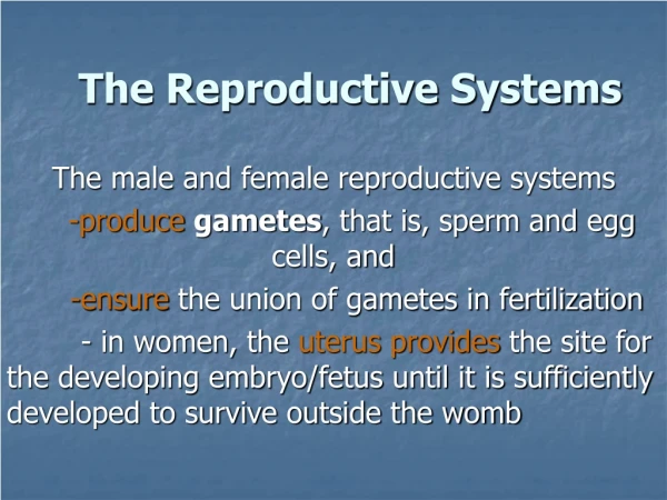
The Reproductive Systems. Sexual reproduction produces new individuals germ cells called gametes (sperm & 2nd oocyte) fertilization produces one cell with one set of chromosomes from each parent Gonads produce gametes & secrete sex hormones Reproductive systems

E N D
The Reproductive Systems • Sexual reproduction produces new individuals • germ cells called gametes (sperm & 2nd oocyte) • fertilization produces one cell with one set of chromosomes from each parent • Gonads produce gametes & secrete sex hormones • Reproductive systems • gonads, ducts, glands & supporting structures • Gynecology is study of female reproductive system • Urology is study of urinary system & male reproductive system
Chromosomes in Somatic Cells & Gametes • Somatic cells (diploid cells) • 23 pairs of chromosomes for a total of 46 • each pair is homologous since contain similar genes in same order • one member of each pair is from each parent • 22 autosomes & 1 pair of sex chromosomes • sex chromosomes are either X or Y • females have two X chromosomes • males have an X and a smaller Y chromosome • Gametes (haploid cells) • single set of chromosomes for a total of 23 • produced by special type of division: meiosis
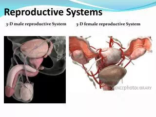
Male Reproductive System • Gonads, ducts, sex glands & supporting structures • Semen contains sperm plus glandular secretions
Formation of Sperm Spermatogenesis is formation of sperm cells from spermatogonia.
Supporting Cells of Sperm Formation • Sertoli cells -- extend from basement membrane to lumen • form blood-testis barrier • support developing sperm cells • produce fluid & control release of sperm into lumen
Spermatogenesis • Spermatogonium (stem cells) give rise to 2 daughter cells by mitosis • One daughter cell kept in reserve -- other becomes primary spermatocyte • Primary spermatocyte goes through meiosis I • DNA replication • tetrad formation • crossing over
Spermatogenesis • Secondary spermatocytes are formed • 23 chromosomes of which each is 2 chromatids joined by centromere • goes through meiosis II • 4 spermatids are formed • each is haploid & unique • all 4 remain in contact with cytoplasmic bridge • accounts for synchronized release of sperm that are 50% X chromosome & 50% Y chromosome
Hormonal Control of Spermatogenesis • Puberty • hypothalamus increases its stimulation of anterior pituitary with releasing hormones • anterior pituitary increases secretion LH & FSH • LH stimulates Leydig cells to secrete testosterone • an enzyme in prostate & seminal vesicles converts testosterone into dihydrotestosterone (DHT-more potent) • FSH stimulates spermatogenesis • with testosterone, stimulates sertoli cells to secrete androgen-binding protein (keeps hormones levels high) • testosterone stimulates final steps spermatogenesis
Hormonal Effects of Testosterone • Testosterone & DHT bind to receptors in cell nucleus & change genetic activity • Prenatal effect is born a male • At puberty, final development of 2nd sexual characteristics and adult reproductive system • sexual behavior & libido • male metabolism (bone & muscle mass heavier) • deepening of the voice
Semen • Mixture of sperm & seminal fluid • glandular secretions and fluid of seminiferous tubules • slightly alkaline, milky appearance, sticky • contains nutrients, clotting proteins & antibiotic seminalplasmin • Typical ejaculate is 2.5 to 5 ml in volume • Normal sperm count is 50 to 150 million/ml • actions of many are needed for one to enter • Coagulates within 5 minutes -- reliquefies in 15 due to enzymes produced by the prostate gland • Semen analysis----bad news if show lack of forward motility, low count or abnormal shapes
Penis • Passageway for semen & urine • Body composed of three erectile tissue masses filled with blood sinuses • Composed of bulb, crura, body & glans penis
Cross-Section of Penis • Corpora cavernosa • upper paired, erectile tissue masses • begins as crura of the penis attached to the ischial &pubic rami and covered by ischiocavernosus muscle • Corpus spongiosum • lower erectile tissue mass • surrounds urethra • begins as bulb of penis covered by bulbospongiosus muscle • ends as glans penis
Root of Penis & Muscles of Ejaculation • Bulb of penis or base of corpus spongiosum enclosed by bulbospongiosus muscle • Crura of penis or ends of corpora cavernosa enclosed by ischiocavernosus muscle
Erection & Ejaculation • Erection • sexual stimulation dilates the arteries supplying the penis • blood enters the penis compressing the veins so that the blood is trapped. • parasympathetic reflex causes erection • Ejaculation • muscle contractions close sphincter at base of bladder and move fluids through ductus deferens, seminal vesicles, & ejaculatory ducts • ischiocavernous & bulbospongiosus complete the job
Female Reproductive System • Ovaries produce 2nd oocytes & hormones • Uterine tubes transport fertilized ova • Uterus where fetal development occurs • Vagina & external genitalia constitute the vulva • Mammary glands produce milk
Follicular Stages • Stages of follicular development • primordial • primary • secondary • graafian • ovulation • Corpus luteum is ovulation wound • fills in with hormone secreting cells • Corpus albicans is white scar left after corpus luteum is not needed
Histology of a Graafian Follicle • Zona pellucida -- clear area between oocyte & granulosa cells • Corona radiata is granulosa cells attached to zona pellucida--still attached to oocyte at ovulation • Antrum formed by granulosa cells secreting fluid • By this time, the oocyte has reached the metaphase of meiosis II stage and stopped developing -- first polar body has been discarded
Life History of Oogonia • Germ cells from yolk sac migrate to ovary & become oogonia • As a fetus, oogonia divide to produce millions by mitosis but most degenerate (atresia) • Some develop into primary oocytes & stop in prophase stage of meiosis I • 200,000 to 2 million present at birth • 40,000 remain at puberty but only 400 mature during a woman’s life • Each month, hormones cause meiosis I to resume in several follicles so that meiosis II is reached by ovulation • Penetration by the sperm causes the final stages of meiosis to occur
Histology of the Uterus • Endometrium • simple columnar epithelium • stroma of connective tissue and endometrial glands • stratum functionalis • shed during menstruation • stratum basalis • replaces stratum functionalis each month • Myometrium • 3 layers of smooth muscle • Perimetrium • visceral peritoneum
Blood Supply to the Uterus • Uterine arteries branch as arcuate arteries and radial arteries that supply the myometrium • Straight & spiral branches penetrate to the endometrium • spiral arteries supply the stratum functionalis • their constriction due to hormonal changes starts menstrual cycle
Vulva (pudendum) • Mons pubis -- fatty pad over the pubic symphysis • Labia majora & minora -- folds of skin encircling vestibule where find urethral and vaginal openings • Clitoris -- small mass of erectile tissue • Bulb of vestibule -- masses of erectile tissue just deep to the labia on either side of the vaginal orifice
Female Reproductive Cycle • Controlled by monthly hormone cycle of anterior pituitary, hypothalamus & ovary • Monthly cycle of changes in ovary and uterus • Ovarian cycle • changes in ovary during & after maturation of oocyte • Uterine cycle • preparation of uterus to receive fertilized ovum • if implantation does not occur, the stratum functionalis is shed during menstruation
Hormonal Regulation of Reproductive Cycle • GnRH secreted by the hypothalamus controls the female reproductive cycle • stimulates anterior pituitary to secrete FSH & LH • FSH initiates growth of follicles that secrete estrogen • estrogen maintains reproductive organs • LH stimulates ovulation & promotes formation of the corpus luteum which secretes estrogens, progesterone, relaxin & inhibin • progesterone prepares uterus for implantation and the mammary glands for milk secretion • relaxin facilitates implantation in the relaxed uterus • inhibin inhibits the secretion of FSH
Menstrual Phase • Menstruation lasts for 5 days • First day is considered beginning of 28 day cycle • In ovary • 20 follicles that began to develop 6 days before are now beginning to secrete estrogen • fluid is filling the antrum from granulosa cells • In uterus • declining levels of progesterone caused spiral arteries to constrict -- glandular tissue dies • stratum functionalis layer is sloughed off along with 50 to 150 ml of blood
Preovulatory Phase • Lasts from day 6 to 13 (most variable timeline) • In the ovary (follicular phase) • follicular secretion of estrogen & inhibin has slowed the secretion of FSH • dominant follicles survives to day 6 • by day 14, graafian follicle has enlarged & bulges at surface • increasing estrogen levels trigger the secretion of LH • In the uterus (proliferative phase) • increasing estrogen levels have repaired & thickened the stratum functionalis to 4-10 mm in thickness
Ovulation • Rupture of follicle & release of 2nd oocyte on day 14 • Cause • increasing levels of estrogen stimulate release of GnRH which stimulates anterior pituitary to release more LH • Corpus hemorrhagicum results
Signs of Ovulation • Increase in basal body temperature • Changes in cervical mucus • Cervix softens • Mittelschmerz---pain
Postovulatory Phase • Most constant timeline = lasts 14 days • In the ovary (luteal phase) • if fertilization did not occur, corpus albicans is formed • as hormone levels drop, secretion of GnRH, FSH & LH rise • if fertilization did occur, developing embryo secretes human chorionic gonadotropin (hCG) which maintains health of corpus luteum & its hormone secretions • In the uterus (secretory phase) • hormones from corpus luteum promote thickening of endometrium to 12-18 mm • formation of more endometrial glands & vascularization • if no fertilization occurs, menstrual phase will begin