
Joints: Classification, Types, and Functions
E N D
Presentation Transcript
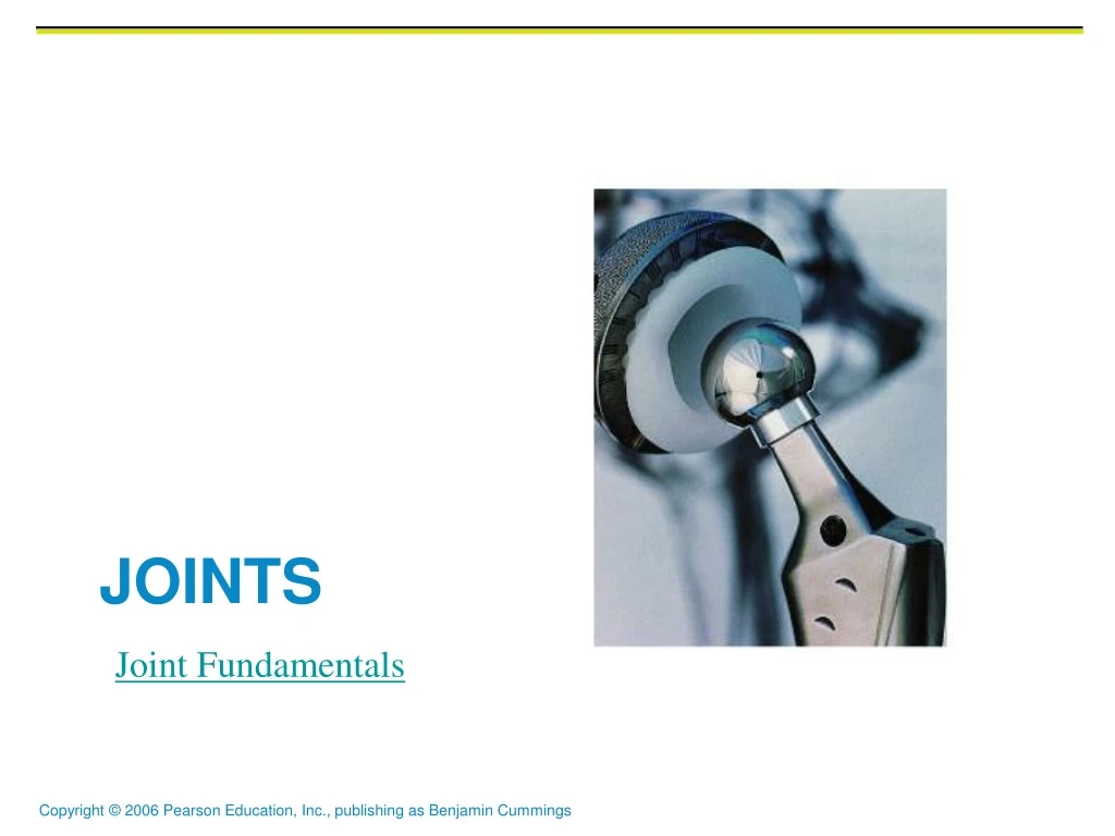
Joints Joint Fundamentals
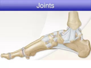
Joints (Articulations) • Weakest parts of the skeleton • Articulation – site where two or more bones meet • Functions of joints • Give the skeleton mobility • Hold the skeleton together
Classification of Joints: Structural • Structural classification focuses on the material binding bones together and whether or not a joint cavity is present • The three structural classifications are: • Fibrous • Cartilaginous • Synovial
Classification of Joints: Functional • Functional classification is based on the amount of movement allowed by the joint • The three functional classes of joints are: • Synarthroses – immovable • Amphiarthroses – slightly movable • Diarthroses – freely movable
Fibrous Structural Joints • The bones are joined by dense, connective fibrous tissues • There is no joint cavity • Most are immovable • There are three types – sutures, syndesmoses, and gomphoses
Fibrous Structural Joints: Sutures • Occur between the bones of the skull • Comprised of interlocking junctions completely filled with connective tissue fibers • Bind bones tightly together, but allow for growth during youth • In middle age, skull bones fuse and are called synostoses
Fibrous Structural Joints: Sutures Figure 8.1a
Fibrous Structural Joints: Syndesmoses • Bones are connected by a fibrous tissue ligament • Movement varies from immovable to slightly variable • Examples include the connection between the tibia and fibula, and the radius and ulna
Fibrous Structural Joints: Syndesmoses Figure 8.1b
Review • Name two functions of joints. • Name three structural classification of joints. • Name three functional classification of joints. • Name three types of fibrous joints. • Where is the most prominent place to find suture joints. • Name one example of Syndesmoses joint. Joint Fundamentals
Fibrous Structural Joints: Gomphoses • The peg-in-socket fibrous joint between a tooth and its alveolar socket • The fibrous connection is the periodontal ligament
Cartilaginous Joints • Articulating bones are united by hyaline cartilage or disks of fibrocartilage • Lack a joint cavity • Two types – synchondroses and symphyses
Cartilaginous Joints: Synchondroses • A bar or plate of hyaline cartilage unites the bones • Examples include: • Epiphyseal plates of children • Joint between the costal cartilage of the first rib and the sternum
Cartilaginous Joints: Synchondroses Figure 8.2a, b
Cartilaginous Joints: Symphyses • Hyaline cartilage covers the articulating surface of the bone and is fused to an intervening pad of fibrocartilage • Amphiarthrotic joints designed for strength and flexibility • Examples include intervertebral joints and the pubic symphysis of the pelvis
Cartilaginous Joints: Symphyses Figure 8.2c
Synovial Joints • Those joints in which the articulating bones are separated by a fluid-containing joint cavity • Ends of the bones are covered in hyaline cartilage and the ends are encapsulated by synovial fluid • All are freely movable • Examples – all limb joints, and most joints of the body
Synovial Joints: General Structure • Synovial joints all have the following • Articular cartilage • Joint (synovial) cavity • Articular capsule • Synovial fluid • Reinforcing ligaments
Synovial Joints: General Structure Figure 8.3a, b
Ball-and-Socket Joints Types of Synovial Joints • A spherical or hemispherical head of one bone articulates with a cuplike socket of another • Multiaxial joints permit the most freely moving synovial joints • Examples: shoulder and hip joints
Ball-and-Socket Joints Figure 8.7f
Condyloid Joints • Oval articular surface of one bone fits into a complementary depression in another • Both articular surfaces are oval • Biaxial joints permit all angular motions • Examples: radiocarpal (wrist) joints, and metacarpophalangeal (knuckle) joints
Condyloid Joints Figure 8.7d
Planar Joints • One flat bone surface glides or slips over another similar surface • Examples – intercarpal and intertarsal joints, and between the flat articular processes of the vertebrae
Planar Joints Figure 8.5a
Hinge Joints • Hinge joints • Cylindrical projections of one bone fits into a trough-shaped surface on another • Motion is along a single plane • Uniaxial joints permit flexion and extension only • Examples: elbow and interphalangeal joints
Hinge Joints Figure 8.7b
Pivot Joints • Rounded end of one bone protrudes into a “sleeve,” or ring, composed of bone (and possibly ligaments) of another • Only uniaxial movement allowed • Examples: joint between the axis and the dens, and the proximal radioulnar joint
Pivot Joints Figure 8.7c
Saddle Joints • Similar to condyloid joints but allow greater movement • Each articular surface has both a concave and a convex surface • Example: carpometacarpal joint of the thumb Joints
Saddle Joints Figure 8.7e