Download
1 / 12
130 likes | 398 Vues
Flow Cytometric Analysis of FRET to Study the Interaction Between CFP- and YFP-Tagged Proteins. David Stepensky. Classical pathway of major histocompatibility complex (MHC) class I antigen processing, loading, and presentation. Groothuis et al, Immunol Rev 2005. Objectives.

E N D
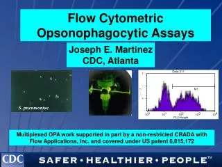
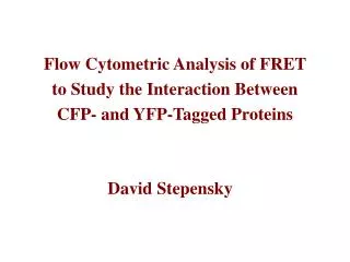
Flow Cytometric Analysis of FRET to Study the Interaction Between CFP- and YFP-Tagged Proteins David Stepensky
Classical pathway of major histocompatibility complex (MHC) class I antigen processing, loading, and presentation Groothuis et al, Immunol Rev 2005
Objectives to study the interactions within the MHC I loading complex using fluorescently-tagged components: • the kinetics and sequence of association and dissociation of the loading complex • effects of the individual interactions on the loading of the MHC class I molecules with peptides Cresswell et al, Immunol Rev 2005 Approach • generation of fluorescently tagged components of the loading complex • investigation of functioning of the tagged proteins fluorescence-based techniques (FRET, FRAP, etc.) biochemical techniques
Fluorescence Resonance Energy Transfer transfer of excited state energy from one fluorophore to another CFP HC Tapasin CFP YFP Excitation & emission spectra YFP Tapasin HC CFP YFP CFP excitationCFP & FRET signals YFP excitationYFP signal extent of interaction %FRET
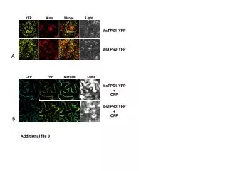
Experimental setup experimental cell lines controls • interaction between MHC class I HC & Tapasin • M553 tapasin deficient melanoma • stable transfection with: • Tapasin-YFP & • MHC I heavy chain-CFP • multiclonal cell lines • interaction (FRET efficiency) was measured using confocal microscope (n=15-20 cells) FRET, % Tapasin-YFP Tapasin C95A-YFP Tapasin-YFP Tapasin C95A-YFP Tapasin-YFP Tapasin C95A-YFP w/o Tapasin w/o Tapasin Tapasin Tapasin-YFP Tapasin C95A-YFP HLA-B44-CFP HLA-A2.1- CFP HLA-A2.1-YFP-CFP HLA-A2.1-CFP HLA-A2.1 T134K-CFP
Flow Cytometric Analysis of FRET Objective: to obtain statistically robust measurement of FRET efficiency in the studied cell lines CFP • FACSAria • Violet laser 405 nm FACS setup: Excitation & emission spectra CFP (450/40 nm) FRET(530/30 nm) YFP • Blue laser 488 nm YFP (530/30 nm) • one laser at a time, sequential acquisition of the same sample 405 CFP 488 FRET/YFP
FACS-FRET: the raw data Negative control Exper. sample FRET CFP Positive control positive control experimental sample negative control FRET cells CFP YFP
FACS-FRET: the results • FACS-FRET results are consistent with the confocal data • both techniques seem to quantify correctly the interaction between the constructs experimental cell lines controls Tapasin-YFP Tapasin C95A-YFP Tapasin-YFP Tapasin C95A-YFP Tapasin-YFP Tapasin C95A-YFP w/o Tapasin w/o Tapasin Tapasin Tapasin-YFP Tapasin C95A-YFP FRET/CFP ratio, normalized HLA-B44-CFP HLA-A2.1- CFP HLA-A2.1-YFP-CFP HLA-A2.1-CFP HLA-A2.1 T134K-CFP
FRET assessment using FACS or confocal microscope: selected characteristics
Alternative setups for FACS-FRET He et al, Cytometry Part A, 2003 Dye, Clin Appl Immunol Rev, 2005 • FACSVantage SE • spatial separation of the laser lines • optional laser • nonstandard mirrors/filters • FACSVantage SE • laser tuning to 458 nm • simultaneous excitation of CFP & YFP • nonstandard mirrors/filters
FACS Analysis of FRET • simple setup • combination of FACS with Confocal analysis • possibility of cell sorting
Thanks Prof. Peter Cresswell and the group Cell Sorter Facility Geoff LyonTom TaylorDon Foster