Download
1 / 50
510 likes | 712 Vues
Head (Brain) Injury. This Session. Skull CSF Circulation Brain stem Head injury Assessment. Structure of the Skull. Bones fused O penings allow passage of blood vessels and nerves L argest opening is the foramen magnum. Production and Circulation of Cerebrospinal Fluid (CSF).

E N D
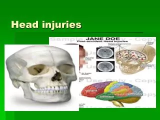
This Session • Skull • CSF • Circulation • Brain stem • Head injury • Assessment
Structure of the Skull • Bones fused • Openings allow passageof blood vessels and nerves • Largest opening is the foramen magnum
Production and Circulation of Cerebrospinal Fluid (CSF) • Production from choroid plexus • One in each ventricle • CSF provides buoyancy, protection & chemical stability to Brain
Production and Circulation of CSF • CSF circulates through the subarachnoid spaces and ventricles • CSF is reabsorbed from the arachnoid villi into the Sagittal Sinus
Circulation of the Brain • Arterial supply (arterial pressure) • Venous sinuses and veins
Brain Stem • Respiratory centres • Cardiac centres • Cranial nerves • Reticular Activating System
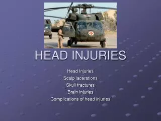
Head Injury Risk • Males, ages 15 to 30 years • Low/median income • Peak- evenings, nights weekends • Alcohol • Causes: motor vehicle accident, falls, assaults, sport-related injuries
Injury • Primary injury – from the impact • Secondary injury – hypoxia, hypercapnia, hypotension (ischaemia), intracranial hypertension (high ICP) • Acceleration-deceleration • Rotational injuries
Injury • Primary Injury • Direct tissue damage from traumatic mechanism (eg. Contusion, tissue shearing, haemorrhage) • Secondary injury • Occurs minutes to hours after the primary injury • Ischemia from elevated ICP and/or systemic hypotension • Metabolic toxins
Open Head Injury • Fractures associated with Open Head Injury • Depressed • Open • Comminuted • Basilar -CSF leakage from nose and ears (i.e. rhinorrhea and otorrhea) • Most often from bullet or knife wounds
Closed Head Injury • Caused by ‘blunt’ trauma • Concussion • Contusion • Laceration • Acceleration & Deceleration Injuries
Concussion • Mild Concussion – cortical dysfunction • Classic Concussion – loss consciousness • No physical evidence
Cerebral Contusion • Cortical bruise • Usually due to violent anterior-posterior displacement • Contusion at point of contact is “coup” • Contusion opposite is “contre coup”
Brain Stem Injury • Poor prognosis • Immediate dysfunction, loss of consciousness, pupillary changes, posturing, cranial nerve deficits, changes in vital functions
Diffuse Axonal Injury • Shear damage is microscopic • Common cause of brain damage after Traumatic Brain Injury
How things evolve after a Head Injury • Brain is enclosed • Blood, brain tissue and CSF contribute to ICP • Normal = 10 - 15 mm Hg in lateral ventricles • Brain Oedema, Brain tumour or bleeding increases ICP
Monroe Kellie Hypothesis BRAIN 80% BLOOD 10% CSF 10% 80% 10% 10%
As ICP increases • Initially compensated by displacement of CSF • ICP increases ⇒ cerebral blood flow decreases ⇒ tissue hypoxia ⇒ decrease in pH and increase in CO2 level • This leads to cerebral vasodilation, oedema & further increases in ICP. This cycle continues
As ICP increases • CNS ischaemic response • Ultimately, brain can herniate
Signs and Symptoms of Increased ICP • LOC change • Pupil change • Motor dysfunction • Headache • Change in Breathing Pattern
Signs & Symptoms of Increased ICP • Vomiting • Positive Babinski reflex • Blurred vision, diplopia • Seizures • Loss of brain stem reflexes • Cushing reflex
Treatment • Reduce ICP surgically or with mannitol (osmotic diuretic)
Epidural Subdural Intracerebral Types ofHaematoma
Epidural Haematoma • Between skull & dura • Usually due to torn middle meningeal artery • Skull usually fractured • Dura slowly separates • Arterial bleed
Intracerebral Haematoma • 2/3 ruptured aneurysms • Also caused by penetrating injuries • CSF often contains blood • Rapid progression as arterial bleed • More frequent in older persons and alcoholics
Subdural Haematoma • Between dura and arachnoid • Common in victims of child abuse • Dura attached to skull, pia to Brain • Bridging veins shear • Bleeding is slower than epidural
Subdural Haematoma • Absence of blood in CSF doesn’t negate subdural haematoma • Clinically manifest as: • Acute • Chronic
Acute & Chronic Subdural Haematomas • Based on time interval until appearance of symptoms after injury • Acute (within 24 hr) • Subacute (2-10 days) • Chronic (possible weeks)
Acute Subdural Haematoma • Symptoms seen within 24 hours • Progresses rapidly and carry high mortality due to secondary injuries from inc. ICP • Similar symptoms to Epidural haematoma due to ICP
Chronic Subdural Haematoma • Develop weeks after injury • Clot is encapsulated by fibroblasts • Encapsulated cells gradually lyse and the contained fluid develops high osmotic pressure • Draws fluid from surrounding tissue, increasing volume (& ICP)
Chronic Subdural Haematoma • Affects older persons with cerebral atrophy • Minor fall causes subdural haemorrhage - often subclinical • Clot liquification over next 2-4 weeks results in a process of clot expansion and development of signs and symptoms of a mass • Effects may resemble brain tumour or stroke
Chronic Subdural Haematoma • Treatment usually surgical • Most patients make excellent recovery unless elevated ICP leads to secondary injury or herniation
Characterization of TBI • Clinical severity is graded using GCS • Mild, GCS 13-15 • normal to lethargic, mildly disoriented • Moderate, GCS 9-12 • lethargic to obtunded, follows commands with arousal, confused • Severe, GCS 3-8 • comatose, no eye opening or verbalization. • does not follow commands • motor exam: ranges from localizing to posturing
Glasgow Coma Scale Eye Opening Response • Spontaneous--open with blinking at baseline 4 points • To verbal stimuli, command, speech 3 points • To pain only (not applied to face) 2 points • No response 1 point Verbal Response • Oriented 5 points • Confused conversation, but able to answer questions 4 points • Inappropriate words 3 points • Incomprehensible speech 2 points • No response 1 point Motor Response • Obeys commands for movement 6 points • Purposeful movement to painful stimulus 5 points • Withdraws in response to pain 4 points • Flexion in response to pain (decorticate posturing) 3 points • Extension response in response to pain (decerebrate posturing) 2 points • No response 1 point