TRAUMA TO THE HEAD
170 likes | 415 Vues
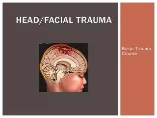
TRAUMA TO THE HEAD. TRAUMA TO THE SCALP (LACERATIONS) 2.TRAUMA TO THE SCALP ( FRACTURES) 3.TRAUMA TO BRAIN CONTUSIONS LACERATIONS HEMORRHAGE Prof. C. E. Connolly. SCALP.

TRAUMA TO THE HEAD
E N D
Presentation Transcript
TRAUMA TO THE HEAD TRAUMA TO THE SCALP (LACERATIONS) 2.TRAUMA TO THE SCALP ( FRACTURES) 3.TRAUMA TO BRAIN CONTUSIONS LACERATIONS HEMORRHAGE Prof. C. E. Connolly
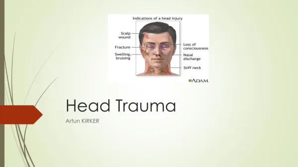
SCALP • Because of tight appostion of the scalp to the calavrium – lacerations easily occur • Bleed copiously +++ Hard to stop. There is free communication between the vessels of the scalp and face to the veins of the meninges. • Danger of Meningitis especially when the laceration is accompanied by a # of skull. There may be grave injury to the brain without skull # or scalp injury.
Fractures of the Skull • 1. Localised Depressed Fracture: due to blunt force .ie. Hammer blow. Plank of wood etc. • 2. Linear Fracture due to R.T.A. Blunt force or a simple fall often to the Base of skull ( easy to miss on X Ray) • Note • Look for bleeding from the ear(s) or into the orbit – Black Eyes- Hematuma of Eye - # Base of skull. Leak of C.S.F. from nose – Clear Fluid.
Concussion/ Loss of Consciousness Injury • Instantaneous loss of function of loss of consciousness followed by rapid (mins) and complete recovery. • If consciousness lost the individual experiences no sensation until his sudden rather surprised awakening. • Retrograde Amnesia- No memory of blow. Duration of loss of consciousness is a guide to the degree of cerebral pathology. • Pathology Mild degrees of Diffuse Axonal Injury ( D.A.I.)
Intracranial Hemorrhage • 1. Extradural: Blood between Bone and (“Epidural”) Dura. 2.Subdural: Blood between Dura and Brain. 3. Subarachnoid: Blood beneath the Leptomeninges due to a ruptured Berry Aneurysm or Trauma – RTA or Blow to side fo the upper neck ( Karate Chop!)
Extradural • Clinical Trauma (Kick, Blow) to the side of the head • Concussion – Rapid Recovery- Lucid interval- Loss of consciousness – coma- Death with 6-12 Hours or less! • Path # of Temporal Bone (Fragile) with tearing of the middle meningeal artery/vein Slow bleeding with gradual separation of Dura from Bone over period of 6-12 Hours . Accumulation of a large Hematoma • Outside Dura • Compression of Brain.
Subdural • Clinical : Elderly Patient – Minor trauma (fall etc.) to head . Usually no Skull fracture. Presents a week or two later with C.N.S Deficit i.e. Memory loss, Blurred vision , Headache, Epilepsey • PATHOLOGY • Tearing of Veins as they enter the Sup. Saggital Sinus due to the shearing force on veins coming from a small atrophic brain which is oscillating due to minor trauma • Slow Venous Oozing into subdural space • (200 – 500 mls) • Granulation tissue grows into Hematoma from Dura.
Subdural • Path Hematoma becomes encapsulated by granulation tissue – thin capsule formed. Hematoma may increase in volume by • 1. Rebleeding from granulation tissue • 2. Hyerosmotic state Hematoma may draw in CSF from subarachnoid space below • 3. Further falls, trauma etc.
Head Trauma - Children • Child’s skull bones are pliable. Unusual to see fractures. Usually see Ping-Pong Ball indentations ( “fractures”) in skull . • Middle Meningeal Artery torn much less often. • Separation of sutures often seen with violent trauma rather than a fracture. • Tearing of Bridging veins from Cerebral Cortex to Sup. Saggital Sinus Acute Subdural Hematoma. I.C.P.
Coup Injuries • When the stationary head is struck with a blunt instrument (i.e hammer) contusions are located beneath the point of impact • Contusion- Pinpoint Necrosis of Brain Tissue – rupture of tiny capillaries – Bleeding resolution over weeks –months – Tiny Brown concave depression.
ContraCoup Injuries • When the moving head strikes a firm surface. i.e. (Footpath, Road, etc.) Brain contusions are located opposite the point of impact in the absence of a skull fracture FALL FALL
Contra Coup ( Mechanism) • Brain Lag: • As the skull is accelerated towards the ground the brain will lag towards the anterior surface compared to the CSF insulation fluid which shifts immediately in the direction of CSF • Acceleration sloshs to the back. Fails to insulate brain anteriorly – Damaged against underlying rough projection bone.
Trauma to the Head • 1. Blunt trauma due to blows to Head • Laceration of Scalp • Fractures of Skull • Extra-Dural Hematoma – Contusions of Brain 2. Head-in-Motion Injury Falls, RTA’s, Boxers, etc. Subdural Hematoma (No fractures) (Tearing of veins) AND/OR Diffuse Axonal Injury (D.A.I) (Shearing of Axons) (90% due to RTA’s)
Acute Traumatic Subarchnoid Hemorrhage • Cause: Trauma to the Verteral Artery with a Laceration. Artery is most vulnerable on the transverse process of C1 where it emerges from the foramina Bleeding under pressure into the subarachnoid space up. In the posterior fossa. • Trauma may be due to • 1. Blow to the side of the behind the Ear (Karate Chop) • 2. Acute Rotational Movement to the Head
Trauma to the Brain . II Children • Commonest cause of Death in Children – Intracranial Hemeorrhage + Skull Fracture • Specifically • Subdural Hematoma Blunt Impact ( Fist , Fall etc.) • Cause Shaking Head (“Shaking Baby Syndrome”) • Death due to Cerebral Edema ++++ Diffuse Axonal Damage Sub Dural Haem. due to rupture of vessels in subdural space.