Chapter 9 Joints
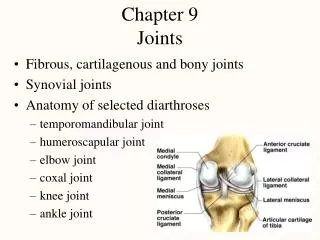
Chapter 9 Joints. Fibrous, cartilagenous and bony joints Synovial joints Anatomy of selected diarthroses temporomandibular joint humeroscapular joint elbow joint coxal joint knee joint ankle joint. Joints and Their Classification. Arthrology is the study of the joints

Chapter 9 Joints
E N D
Presentation Transcript
Chapter 9Joints • Fibrous, cartilagenous and bony joints • Synovial joints • Anatomy of selected diarthroses • temporomandibular joint • humeroscapular joint • elbow joint • coxal joint • knee joint • ankle joint
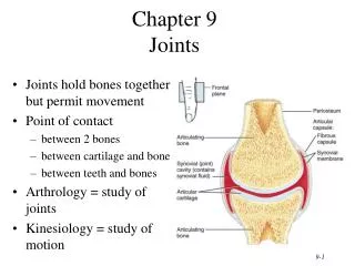
Joints and Their Classification • Arthrology is the study of the joints • Kinesiology is the study of musculoskeletal movement • Joints are classified by their freedom of movement • diarthrosis (freely movable); amphiarthrosis (slightly movable) and synarthrosis (little or no movement) • Joints are classified by the manner adjacent bones are joined -- fibrous, cartilaginous, bony and synovial joints
Fibrous, Cartilaginous & Bony Joints • Fibrous joints have collagen fibers spanning the space between bones • sutures, gomphoses & syndesmoses • Cartilaginous joints have 2 bones bound to each other by cartilage • synchondroses or symphyses • Bony joints have 2 bones fused by osseous tissue • synostoses in early adulthood
Fibrous Joint -- Sutures • Immovable fibrous joints that bindthe bones of the skull to each other • Serrate sutures appear as interlocking wavy lines • coronal, sagittal & lambdoid sutures • Lap or squamous sutures are 2 bones with overlapping beveled edges • temporal & parietal bones • Plane or butt sutures have straight, nonoverlapping edges • palatine processes of the maxillae
Fibrous Joint -- Gomphoses • Attachment of a tooth to itssocket is a joint called agomphoses • Tooth held in place by fibrous periodontal ligament • collagen fibers that extend from bone of jaw to tooth • Allows tooth to move a little while chewing
Fibrous Joint -- Syndesmoses • Joint in which two bones are bound by a ligament only (interosseus membrane) • Most movable of fibrous joints • Interosseus membranes unite radius to ulna and tibia to fibula
Cartilaginous Joint -- Synchondroses • Bones are joined by hyaline cartilage • rib attachment to sternum by • epiphyseal plate in children binds epiphysis and diaphysis
Cartilaginous Joint -- Symphyses • 2 bones joined by fibrocartilage • pubic symphysis and intervertebral discs • Only slight amount of movement is possible
Bony Joints (Synostoses) • 2 bones, once separate, fused by osseous tissue • Ossification occurs with age • left and right mandible present at birth • left and right frontal bones present at birth • epiphyses and diaphysis of the long bones
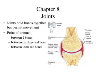
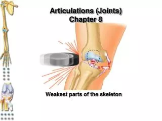
Synovial Joint • Joint in which two bones are separated by a space called a joint cavity • Most are freely movable
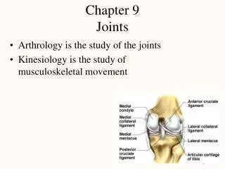
General Anatomy of Synovial Joints • Articular capsule • fibrous capsule lined by synovial membrane • continuous with periosteum • Synovial fluid • viscous slippery fluid rich in albumin & hyaluronic acid & similar to raw egg white • Articular cartilage • hyaline cartilage covering the joint surfaces • Meniscus is pad of fibrocartilage injaw, wrist, knee and sternoclavicular joints • absorbs shock, guides bone movements & distributes forces • Tendon attaches muscle to bone • Ligament attaches bone to bone
Tendon Sheaths and Bursae • Bursa is saclike extension of joint capsule that extends between nearby structures allowing them to slide more easily past each other • Tendon sheaths are elongated cylinders of connective tissue lined with synovial membrane & wrapped around a tendon • numerous in hand and foot
Ball-and-Socket Joints • Smooth hemispherical head fits within a cuplike depression • head of humerus into glenoid cavity of scapula • head of femur into acetabulum of hip bone • Multiaxial joint
Hinge Joints • One bone with convex surface that fits into a concave depression on other bone • ulna and humerus at elbow joint • femur and tibia at knee joint • finger and toe joints • Monoaxial joint
Saddle Joints • Each articular surface is shaped like a saddle, concave in one direction and convex in the other • trapeziometacarpal joint at the base of the thumb • Biaxial joint • more movable than a condyloid or hinge joint forming the primate opposable thumb
Pivot Joints • One bone has a projection that fits into a ringlike ligament of another • First bone rotates on its longitudinal axis relative to the other • atlantoaxial joint (dens and atlas) • proximal radioulnar joint allows the radius during pronation and supination
Gliding Joints • Flat articular surfaces in which bones slide over each other • Limited monoaxial joint • Considered amphiarthroses
Condyloid (ellipsoid) Joints • Oval convex surface on one bone fits into a similarly shaped depression on the next • radiocarpal joint of the wrist • metacarpophalangeal joints at the bases of the fingers • Biaxial joints
Flexion, Extension & Hyperextension • Flexion decreases the angle of a joint • bending elbow or wrist • Extension straightens a joint and returns a body part to the anatomical position • Hyperextension is extension of a joint beyond 180 degrees
Abduction & Adduction • Abduction is movement of a part away from the midsagittal line -- raising the arm to the side • Adduction is movement towards the midsagittal line
Abduction & Adduction • Abduction is spreading the fingers away from the midline (middle finger) • Adduction is movement is returning the fingers to the anatomical position
Elevation and Depression • Elevation is a movement that raises a bone vertically • mandibles are elevated during biting & clavicles during a shrug • Depression is lowering the mandible or the shoulders
Protraction & Retraction • Protraction is movement of a bone anteriorly (forward) on a horizontal plane • thrusting the jaw forward, shoulders or pelvis forward • Retraction is movement of a bone posteriorly
Lateral & Medial Excursion • Lateral excursion is sideways movement to right or left • Medial excursion is movement back to the midline • Side-to-side grinding movements occurring during chewing
Circumduction • Movement in which one end of an appendage remains stationary while the other end makes a circular motion • Sequence of flexion, abduction, extension & adduction movements • baseball player winding up for a pitch
Lateral and Medial Rotation • Movement of a bone turning on its longitudinal axis • rotation of trunk, thigh, head or arm • Medial rotation turns the bone inwards • Lateral rotation turns the bone outwards
Supination & Pronation • Occurs in the forearm and foot • Supination • rotation of forearm so that the palm faces forward • inversion and abduction of foot (raising the medial edge of the foot) • Pronation • rotation of forearm so the palm faces to the rear • eversion and abduction of foot (raising the lateral edge of the foot)
Opposition & Reposition • Opposition is movement of the thumb to approach or touch the fingertips • Reposition is movement back to the anatomical position • Important hand function that enables the hand to grasp objects
Dorsiflexion & Plantar Flexion • Dorsiflexion is raising of the toes as when you swing the foot forward to take a step (heel strike) • Plantarflexion is extension of the foot so that the toes point downward as in standing on tiptoe
Inversion & Eversion • Inversion is a movement in which the soles are turned medially • Eversion is a turning of the soles to face laterally
Range of Motion • Varies greatly from one type of joint to another • Measured with goniometer • Factors affecting ROM and joint stability • structure & action of themuscles • proprioceptors keep track of joint position & muscle tone • structure of the articular surfaces • strength and tautness of ligaments, tendons & capsule • gradual stretching of ligaments increases range of motion • “double-jointed” people have unusually long or slack ligaments
Components of a Lever • A lever is a rigid object that rotates around a fixed point called a fulcrum • Rotation occurs when effort overcomes resistance • resistance arm & effort arm are described relative to fulcrum
Mechanical Advantage of a Lever • Ratio of output force to input force ( for math see next slide) • Why would a joint have 2 or more muscles acting on it? • insertions of muscles are slightly different with different mechanical advantages • runner uses high mechanical advantage muscles to overcome body’s inertia & start moving • runner shifts to muscles that have lower MA but produce more speed • Architecture of muscular system has a purpose
Mechanical Advantage • Mechanical advantage is calculated from the length of the effort arm divided by the length of the resistance arm • Contraction of the biceps muscle causes the hand to move quickly • Contraction of the small digastric muscle opens the mouth
First-Class Lever • Has fulcrum in the middle between effort & resistance • Atlantooccipital joint lies between the muscles on the back of the neck and the weight of the face • loss of muscle tone occurs when you nod off in class
Second-Class Lever • Has resistance in the middle between fulcrum & effort • Resistance from the muscle tone of the temporalis muscle lies between the jaw joint and the pull of the diagastric muscle on the chin as it opens the mouth quickly • upside down example relative to wheelbarrow illustration
Third-Class Lever • Has the effort in the middle between the resistance & the fulcrum • most joints of the body • The effort applied by the biceps muscle is applied to the forearm between the elbow joint and the weight of the hand and the forearm
Temporomandibular Joint • TMJ syndrome caused by malocclusion & stress • Clicking sounds, headaches, vertigo, pain, or tinnitus
The Humeroscapular Joint • Shoulder is most freely movable joint in the body • shallowness of glenoid & looseness ofcapsule • deepened by glenoid labrum • Supported by ligaments & tendons • 3 glenohumeral, coracohumeral,transverse humeral & biceps tendon areimportant joint stabilizer • Supported by rotator cuff musculature • tendons of 4 muscles form rotator cuff that fuses to joint capsule & strengthens it • supraspinatus, infraspinatus, teres minor & subscapularis, • 4 Bursae associated with shoulder joint
The Elbow Joint • Single joint capsule enclosing the humeroulnar and humeroradial joints • Humeroulnar joint is supported by collateral ligaments. • Radioulnar joint is head of radius held in place by the annular ligament encircling the head
The Hip Joint • Head of femur articulates with acetabulum • Socket deepened by acetabular labrum • transverse acetabular ligament completes labrum • Blood supply to head of femur found in ligament of the head of the femur (round ligament) • Joint capsule strengthened by ligaments
Hip Joint • Joint capsule strengthened by ligaments • pubofemoral • ischiofemoral • iliofemoral