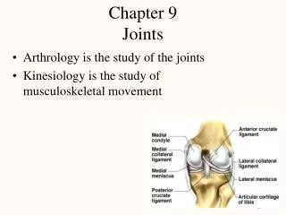
Chapter 9 Joints
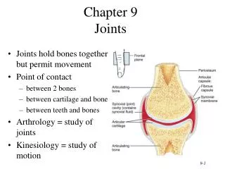
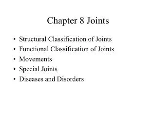
Chapter 9 Joints. Arthrology is the study of the joints Kinesiology is the study of musculoskeletal movement. 2 CLASSIFICATION OF JOINTS. 1. How the bones are joined fibrous, cartilaginous, bony and synovial joints 2. How the bones move We will look at this type of joint mainly.

Chapter 9 Joints
E N D
Presentation Transcript
Chapter 9Joints • Arthrology is the study of the joints • Kinesiology is the study of musculoskeletal movement
2 CLASSIFICATION OF JOINTS • 1. How the bones are joined • fibrous, cartilaginous, bony and synovial joints • 2. How the bones move • We will look at this type of joint mainly
Three types of Joint MOVEMENT • Three ways bones move: • 1. Freely • 2. Slightly • 3. Not at all • A freely moving joint is a DIARTHROSES • This includes All synovial joints • Slightly moving is a AMPHIARTHROSIS • No movement is a SYNARTHROSIS
SYNARTHROSIS • Synarthrosis ( How much does this move?), three types: • 1. Suture • 2. Gomphosis • Teeth • 3. Synchondroses (SYN= together, chrondro= cartilage • Epiphyseal plate
AMPHIARTHROSIS • Amphiarthrosis (moves ________), two types: • 1. Syndesmosis (struck rock twice) • Distal tibiofibular joint • (picture tibula and fibula stricking each other) • 2. Symphyses • Always in the midline of the body • Pubic symphyses • Intervertebral disc
Making Sense of Joints“Cartilage becomes Bone” • A Synchondrosis: 2 bones, once separate, fused by osseous tissue • An example is the epiphyseal plate at the end of growing bones • As the bone grows the synchondrosis (cartilage) is replaced by bone and becomes a synostosis (ost- is bone)
Synarthrosis Joints(Immovable Joint) • Synathrosis • Fibrous joints of collagen fibers in space between bones • sutures, synchondroses, synostosis, gomphosis
Amphiarthrosis(Slightly Moveable Joint) • Symphysis • pubic symphysis and intervertebral discs • Syndesmosis • Distal tibia and fibula joint
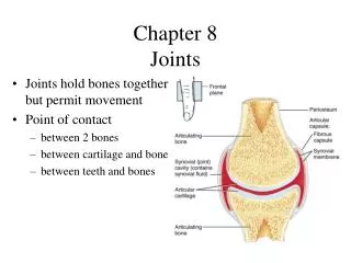
Diarthrosis (Freely Moveable) I • Space between bones called the synovial (joint) cavity • Contains articular capsule and an articular cartilage • 1. Articular capsule is composed of two layers: • 1) the outer fibrous capsule • lined by synovial membrane • continuous with periosteum • 2) the inner synovial membrane • a) which secretes a lubricating and joint-nourishing synovial fluid • viscous slippery fluid rich in albumin & similar to raw egg white • 2. Articular cartilage • hyaline cartilage covering the joint surfaces
Diarthrosis (Freely Moveable) III • Many diarthroses also contain accessory structures: • Extracapsular and intracapsular ligaments • Collateral ligaments of knee • Cruciate ligaments of knee • Articular discs • Meniscus is a pad of fibrocartilage injaw, wrist, knee and sternoclavicular joints • absorbs shock, guides bone movements • Bursae • Found between tendons, muscles, ligaments and bones • Tendon attaches muscle to bone • Ligament attaches bone to bone
Bursae and Tendon Sheaths • Bursa is a sac, an extension of joint capsule that are between nearby structures allowing them to slide more easily past each other • Tendon sheaths are cylinders lined with synovial membrane that wrap around a tendon • numerous in hand and foot • Tenosynovitis: inflammation of the tendon sheaths and synovial membranes • Tendonitis: inflammation of _________.
Ball-and-Socket Joints • Smooth hemispherical head fits within a cuplike depression • head of humerus into glenoid cavity of scapula • head of femur into acetabulum of hip bone • Multiaxial joint
Hinge Joints • One bone with convex surface that fits into a concave depression on other bone • ulna and humerus at elbow joint • femur and tibia at knee joint
Saddle Joints • Each articular surface is shaped like a saddle, concave in one direction and convex in the other • trapeziometacarpal joint at the base of the thumb
Pivot Joints • One bone has a projection that fits into a ringlike ligament of another • First bone rotates on the other • atlantoaxial joint (dens and atlas) • proximal radioulnar joint • the radius during pronation and supination
Gliding Joints • Flat surfaces in which bones glide over each other
Condyloid (ellipsoid) Joints • Oval convex surface fits into a concavity • radiocarpal joint of the wrist • metacarpophalangeal joints at the bases of the fingers
Special Movements of Diarthroses • Flexion decreases the angle of a joint • bending elbow or wrist • Extension increases the angle of a joint • Straighten elbow • Hyperextension is extension of a joint beyond 180 degrees
Abduction & Adduction • Abduction is movement of a part away from the midsagittal line -- raising the arm to the side • Adduction is movement adding to midline
Abduction & Adduction • Abduction is spreading the fingers away from the midline (middle finger) • Adduction is movement is returning the fingers to the anatomical position
Elevation and Depression • Elevation is a movement that raises a bone vertically • mandibles are elevated during biting & clavicles during a shrug • Depression is lowering the mandible or the shoulders
Protraction & Retraction • Protraction is movement of a bone anteriorly (forward) on a horizontal plane • thrusting the jaw forward • scapula • Retraction is movement of a bone posteriorly
Circumduction • Combines flexion, abduction, extension & adduction • baseball player winding up for a pitch • NOT Rotation • (but it looks like it)
Lateral and Medial Rotation • Movement of a bone turning • rotation of trunk, thigh, head or arm • Medial rotation turns the bone inwards (internal rotation) • Lateral rotation turns the bone outwards (external rotation)
Supination/ Pronation • Occurs in the forearm • Supination (get soup) • rotation of forearm so that the palm goes from the posterior to the anterior • Pronation (pour it out) • rotation of forearm so the palm goes from anterior to posterior
Inversion & Eversion • Don’t use supination and pronation for the feet, use: • Inversion • raising the medial edge of the foot • Eversion of foot • lowering the medial edge of the foot (Adam and Eve fell)
Plantar Flexion & Dorsiflexion • The foot does not extend • Dorsiflexion is raising of the toes as when you swing the foot forward to take a step (heel strike) • Plantarflexion is when the toes point downward as in standing on tiptoe (or planting a rose between your toes)
Opposition & Reposition • Opposition is movement of the thumb to approach or touch the fingertips • Reposition is movement back to the anatomical position • Important hand function that enables the hand to grasp objects (us vs. apes)
The Humeroscapular Joint • Shoulder is most freely movable joint in the body • shallowness of glenoid cavity & looseness ofcapsule (golf ball in a shot glass) • Supported by ligaments & tendons • Supported by rotator cuff musculature • supraspinatus, infraspinatus, teres minor & subscapularis (“L” is for lateral rotation and “M” is for medial rotation)
Tendons of Rotator Cuff MusclesWhere is dislocation more likely?
The Pitcher • To throw a baseball a pitcher has to use her shoulder muscles: • 1) To ________ to 10 degrees she will use her supraspinatus muscle • 2) To fully abduct, she will use her _______ muscle • 3) To bring her humerus into lateral rotation she will use her ______ and _______ muscle • 4) And to throw, she will bring her humerus into _____ rotation by using her subscapularis muscle.
The Elbow Joint • Single joint capsule enclosing the humeroulnar and humeroradial joints • Radioulnar and Humeroulnar joint
The Hip Joint • Head of femur articulates with acetabulum • Socket deepened by acetabular labrum • transverse acetabular ligament completes labrum • Blood supply to head of femur found in ligament of the head of the femur (round ligament)
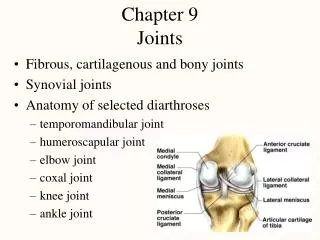
The Knee • Most complex diarthrosis of the body • patellofemoral = gliding joint • tibiofemoral = gliding with slight rotation & gliding possible in flexed position • Joint capsule anteriorly consists of patella & extensions of quadriceps femoris tendon
Knee Joint I • Medial & lateral meniscus absorb shock & shape joint
Knee Joint II • The knee has four major ligaments: two to limit sliding from side to side (collaterals) and front to back (cruciates) • Medial and lateral collateral ligaments prevent rotation of extended knee and side to side slide • Anterior & lateral cruciate ligaments limit anterior & posterior sliding movements (anterior cruciate is on the anterior of the tibia, the posterior cruciate is on the posterior _______. • We use the AP draw test to evaluate these ligament
The Ankle Joint • One joint capsule enclosing the joints between the talus, tibia and fibula • Groups of ligaments • deltoid ligament binding the tibia to the foot on the medial side (never sprained) • lateral collateral ligament binds the fibula to the foot on the lateral side (most ankle sprains) • achilles tendon inserting on the calcaneus
JOINT DISORDERS • Dislocation (luxation): a total displacement of a joint with tearing of ligaments, tendons, and articular capsules (Example: shoulder) • Subluxation: a slight dislocation causing structural and neural dysfunction (Example: spine) • Sprain: ligament wrenching (i.e. sprained ankle) • torn ligaments or tendons • Strain: muscle over stretching
Arthritis • Arthritis is a broad term for pain & inflammation • Blood test may make the diagnosis and/ or X-ray findings • Gouty arthritis • Involves sodium urate crystals deposited in soft tissues of joints, causing inflammation, swelling, and pain • Major features:Great toe, One joint, Uric acid increased, Temperature dependent • Osteoarthritis results from years of joint wear • articular cartilage softens and degenerates • accompanied by crackling sounds called crepitus • bone spurs develop on exposed bone tissue causing pain • Rheumatoid arthritis is autoimmune attack on joint • antibodies attack synovial membrane, enzymes in synovial fluid degrade the cartilage, bones ossify, remissions occur