Equine Skeletal System
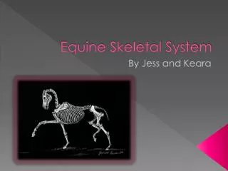
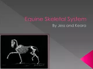
Equine Skeletal System. Axial Skeleton contains bones in the trunk area. Skull Spine (Vertebral column) Ribs Breastbone (Chest cavity) Pelvis Tail. Skull bones – flat, irregular in shape Forms framework for the brain, mouth, eyes & nasal cavity.

Equine Skeletal System
E N D
Presentation Transcript
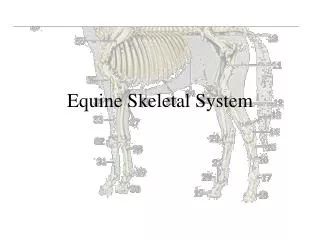
Axial Skeleton contains bones in the trunk area. • Skull • Spine (Vertebral column) • Ribs • Breastbone (Chest cavity) • Pelvis • Tail
Skull bones – flat, irregular in shape Forms framework for the brain, mouth, eyes & nasal cavity. Vertebral column – flexible column of small bones (vertebrae) Hip bones – two large flat bones attached to the spine and sacrum that forms the pelvis or pelvic girdle Ribs & Breastbone (Sternum) along with thoracic vertebrae form the chest cavity
Appendicular Skeleton – forelegs and hindlegs used for locomotion, grooming, defense and feeding • Forelegs – connected to axial skeleton by muscles, not skeletal attachments • Hindlegs – attached to pelvis at the hip joint
Articulations (joints) – the union of two or more bones or cartilages held together by ligaments, tendons, or a tough fibrous capsule • Joints – classified by structure and move ability • Free moving joints contain a joint cavity between the two surfaces, bones covered with smooth cartilage and held together by ligaments
- Only in heart • Muscles – red, lean meat, composes 50% of total body weight. Stimulated to contract or change shape by nerve impulses from the brain, then sends nerve impulses back to brain indicating the degree of contraction so movement is smooth.
Three Basic Muscle Types 1. Smooth muscles (involuntary) • Visceral muscle • Location – Digestion system & Uterus • Prolonged activity w/o fatigue
2. Cardiac Muscle (Involuntary Striated) - Contractions require no nerve stimulus - Rhythmic contractions with no conscious control
3. Striated, Skeletal Muscles – attached to bones of skeletal system either directly or by tendons and act voluntarily. A. Bones serve as levers and muscles move the body voluntary. B. Arranged in opposite sets – one set of muscles bend the limb (flexor muscle), one set straightens the limb (extensor muscle) C. Voluntary muscles become fatigued & need short rest periods.
Tendons – eliminate undue friction, allows muscle to move freely. A. Tendon sheath is a synovial sac through which a tendon passes. Synovial lubricates the surrounding tendon. B. Tendon bursa is a synovial sac interposed between the tendon and the surface over which it comes in contact which lubricates and cushions the tendon. C. Both found near joints
Tendons are fibrous cords of connective tissue attaching muscle to bone, cartilage or other muscle. Tendons insert into bone or cartilage by means of small spikes known as ‘Sharpey’s fibres’.
Functional Anatomy • Flexors bend the limb • Decrease angle of a joint • Examples: Teres major (front leg), Iliacus (hind leg)
Functional Anatomy • Extensors straighten the limb • Increase the angle of a joint • Examples: Brachiocephalicus (front leg), Gluteus medius (hind leg)
Functional Anatomy • Abductors • Move a limb away from the center plane of the equine • Example – Deltoid (front leg)
Functional Anatomy • Adductors • Pulls a limb toward the center plane of the equine • Example: Pectoral muscles (front leg)
Digestive System Digestive system converts feed into a form to use for maintenance, growth and reproduction. Parts – mouth, pharynx, esophagus, stomach, intestines, anus, liver, teeth, pancreas and salivary glands A. Rate of feed passage through stomach – (30 minutes to 2 hours) Feed passes through S. I. Rapidly. Feed not digested and absorbed in the S. I. passes into the cecum (colon) w/n 2 to 4 hours
B. Grinding decreases feed size Increases rate of passage, decreases absorption of nutrients C. Large amounts of concentrates, overwhelming the digestive capacity of the stomach and S.I., can ferment and produce gas or lactic acid causing colic or founder if they pass through the cecum
Mouth extends from lips to the pharynx. Bounded on the sides by cheeks, above by hard palate and below by tongue • Pharynx is muscular, funnel like tube from back of mouth to esophagus directs food and serves as air passage. • Esophagus extends from the pharynx down left side of neck through thoracic cavity and diaphragm to stomach at an angle which makes regurgitation impossible
Stomach – U-shaped muscular sac at front of abdominal cavity. Makes up less than 10% of total digestive capacity for the adult • Small Intestine – 2” in diameter tube. Site of most nutrient absorption. 30% of total digestive capacity.
(1) Anus. (2) Rectum. (3) Base of caecum. (4) Small intestine. (5) Kidney. (6) Liver. (7) Diaphragm. (8) Oesophagus. (9) Large colon. (10) Caecum. (11) Small colon
Large Intestine – consist of cecum, large colon, small colon, rectum and anus • Cecum & Colon 65% of digestive capacity. Enlarged to allow bacteria time to break down large quantities of cellulose • Small colon extends from large colon to the rectum. Place where balls of dung formed. Moisture is reabsorbed in L. I. Creating solid contents