Download
1 / 18
190 likes | 475 Vues
Scanning Probe Microscopy and Nanotechnology for Biomedical Applications. M. Dudziak Silicon Dominion / IEPB May 6, 1999. Contents. What is SPM and why use it in medicine? AFM, STM, LFM, MFM Some applications and results in life sciences & biomaterials research Approaches and methods -

E N D
Scanning Probe Microscopy and Nanotechnology for Biomedical Applications M. Dudziak Silicon Dominion / IEPB May 6, 1999
Contents • What is SPM and why use it in medicine? • AFM, STM, LFM, MFM • Some applications and results in life sciences & biomaterials research • Approaches and methods - • in vitro // fixed • biological // inorganic • Using adaptive learning algorithms and pattern recognition for control and interpretation
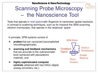
Basics of SPM • Surface forces - Van der Waals, EMF, quantum tunneling • Tip/cantilever movement/current signal • A/D translation of surface reading --> 3D image • Alternative to EM • Partner technology for Optical, Confocal, Near-field Optical Microscopy • Imaging PLUS lithography/fabrication
SPM Technology : 35mm Slides (S) and Overhead Slides (O) • SPM rationale (S) • EM comparisons (S) • STM current/height modes, resolution (S) • AFM principles, types (S, O) • Other Modes (S) • Instrument variation and nonlinearities (S) • Different sample types and images (S) • Sets 0, 1, 2
Research Application Using SPM • Investigation of quantum field effects (QFT) and bioelectromagnetics upon cell topology, structural dynamics, growth • Fundamentally a mathematical, geometrical approach to questions of differentiation and communication • Emphasis upon cytoskeletal and membrane topological features • Search to measure (bio)solitons, fractal & p-adic & chaotic measurables
Foundations • Solitons - stable nonlinear waves • Biosolitons in protein (MT, actin) • Dynamics of MT and IF and effects from EMF, Ca+, other gradients • Intriguing possibilities of the “CA” effect : • neighbors, boundaries, population types • How to study? • Theory and modeling • Computer-based simulation • Experimentation (AFM, confocal, MODE)
Experimentation Goals, Requirements • Living cells • Controlled culture growth • Mechanisms for reproducible sample preparation, gradient application, observation techniques • AFM and AFM+Optical+Confocal best way to go • Main accomplishments (to date): • achieved relative stability in imaging • design of testbed • migration path of image data to modeling/analysis
Neural and Epithelial Imaging • Digital Instruments Nanoscope-III • XR1 Xenepus retinal ganglial cell line • L15 media + embryo extract and fetal calf serum • Relatively rapid death during and after imaging • Multiple rinsing + moisture bath • Bioscope much better than simple fluid cell • Typical XY scan 50 m x 50 m • Typical Z scale 2 m
Set 3 of Overhead Slides • Neural and Epithelial AFM Images
Interpretation, Hypotheses and Theory • Fractal and Chaos Dimensions • Prior interesting observables in large-scale biology (organisms, organs, metabolic rate) • MT structure variations in different pathologies, esp. oncological • Soliton modeling (Dubna, Novosibirsk, ‘93-’96)
Sets 4 & 5 of Overhead Slides • Fractal/Chaos/QB overview • Soliton equations and graphs
Conceptual Formulation • Massive large-scale parallel simulated-annealing type computation in phospholipid membranes • Giving rise to • Soliton-like propagations • Converging to modulation of ion channels and • Amplified effects (QP “pilot wave” principle) in cytoskeletal topology • Effecting changes in cell motility, 3D geometry, and cytoplasmic movement of intracellular components • Giving rise to • Changes in inter-cellular membrane signaling and • Intracellular metabolism and reproduction control
A Geometrical Excursion • Projective Geometry (Pappus, Pascal, Desargues, Klein, Veblen) • Metamorphosis of biological form types from a confluence of simple projections • Path curves, pivot transforms, vortices, and buds • Not magic, just numbers
Path.HTM and Pivot.HTM • Work by N. Thomas (UK)
“Hamilton’s Birds of Prey” • A rare and untamable species • Never photographed in the wild • Sensitive to the touch • Easily camouflaged • Giving rise to speculation about the nature of Geometry and Evolution • Known to inhabit large silicon-based forests
Set 6 of Overhead Slides • Computer simulations of quaternion Julia sets by Tim Stilson
Acknowledgements • Basil Hiley, David Bohm, David Finkelstein • Robert Rosen, Valery Sanyuk, Louis Kaufmann • Hiroomi Umezawa, Karl Pribram, Peter Kugler • Matti Pitkanen, Nick Thomas • Eric Henderson, Tim Stilson • Digital Instruments, Park Scientific Instruments • Many students and assistants • NSF, Jeffress Foundation
References and More • Web resources on SPM: • Start with Digital, Park, Rice, JHU, NCSU, IowaSU • Web Resources on MathBio, QB, BioEM: • Principia Cybernetica and links therefrom • Request from MJD and you may receive, eventually • Explore www.silicond.com/library (No librarian or secretary --- self-service)