SCANNING PROBE MICROSCOPY
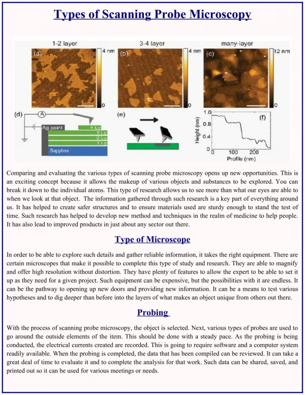
SCANNING PROBE MICROSCOPY. By AJHARANI HANSDAH SR NO.08440. INTRODUCTION. Scanning probe microscopy is an imaging technique in which probe is moved along the surface of specimen. Provides 3D profile of the specimen surface. High resolution imaging technique. DIFFERENT TYPES.


SCANNING PROBE MICROSCOPY
E N D
Presentation Transcript
SCANNING PROBE MICROSCOPY By AJHARANI HANSDAH SR NO.08440
INTRODUCTION • Scanning probe microscopy is an imaging technique in which probe is moved along the surface of specimen. • Provides 3D profile of the specimen surface. • High resolution imaging technique.
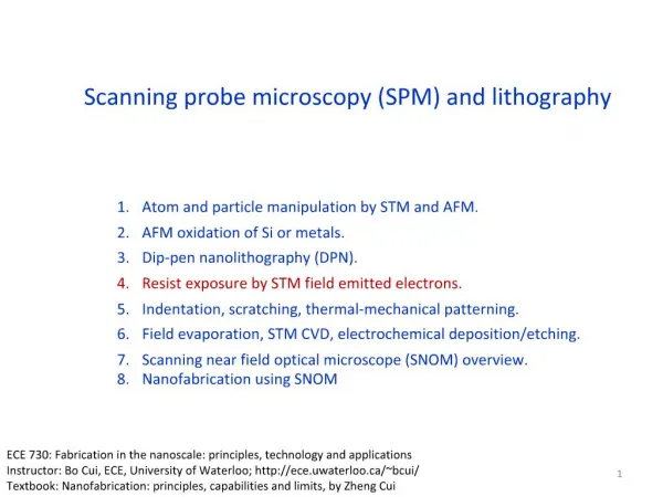
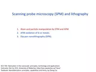
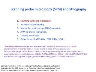
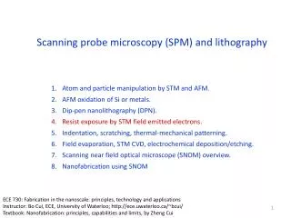
DIFFERENT TYPES • Scanning tunneling microscopy • Atomic force microscopy • Tapping mode AFM • Magnetic force microscopy • Electric force microscopy frictional force microscopy • Near field optical microscopy
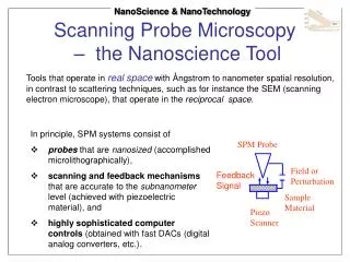
CONT… • Probe is supported on flexible cantilever. • Force depends on stiffness of cantilever and distance between probe and sample surface • Motion of probe over the surface is controlled using feedback loop and piezoelectric scanner • Deflection of probe is measured by beam bounce method • Deflections are used to generate surface topography
MODES OF OPERATION • Contact mode - less than 0.5 nm separation distance between probe and surface. • Tapping mode - 0.5 to 2 nm separation distance. • Non-contact mode - 0.1 to 10 nm separation distance.
CONTACT MODE • Short range interactions. • Spring constant of cantilever is less than surface, hence cantilever bends • Force on tip is repulsive. • Force between probe and sample is constant by feedback loop. • For constant height method - no feed back system is used. • Advantages - fast scanning, good for rough samples, used in friction analysis. • Disadvantages - forces can damage soft samples.
TAPPING MODE • May contact surface. • Cantilever is oscillated at resonant frequency. • Amplitude used as feedback. • Maintaining constant oscillation amplitude, by adjusting tip-sample distance. • Advantages - high resolution images for soft samples, good for biological samples. • Disadvantages - difficult to image in liquids, slower scan speeds.
NON-CONTACT MODE • Attractive Vander Walls forces. • Probe oscillates above surface. • Using a feedback loop to monitor changes in amplitude due to attractive forces. • Advantages - very low force exerted on sample, life of probe is more. • Disadvantage - lower resolution, contaminant layer on surface can interfere with oscillation, usually ultra high vacuum is needed.
ADVANTAGES • 3D surface profile. • Sample does not need any treatments or coatings. • Works well in air or liquid environment. • High resolution.
DISADVANTAGES • Image maximum height of 10-20 micrometers and scanning area of 150x150 micrometers. • Low scanning speed. • Image artifact due to high radius of curvature of tip. • Cannot normally measure steep walls or overhangs.