Download
1 / 61
1.03k likes | 4.25k Vues
SURGICAL ANATOMY OF EXTERNAL AND MIDDLE EAR. Dr Mubeena. Dr Mubeena. Development of external ear. Outer part of first branchial cleft 6 cartilaginous tubercles appear towards the end of first fetal month 3 on first arch 3 on 2 nd 20 th week – adult shape.

E N D
SURGICAL ANATOMY OF EXTERNAL AND MIDDLE EAR Dr Mubeena Dr Mubeena
Development of external ear Outer part of first branchial cleft 6 cartilaginous tubercles appear towards the end of first fetal month 3 on first arch 3 on 2nd 20 th week – adult shape
Development of external auditory canal Ectoderm of first visceral cleft Funnel shaped tube Meatal plug Recanalisation of this plug forms epithelial lining of bony meatus 28th week - fully formed
Development of tympanic membrane Outer epithelial layer – ectoderm of visceral cleft Middle fibrous layer – mesoderm between first visceral cleft and tubotympanic recess Inner mucosal layer – endoderm from a part of recess
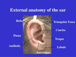
Anatomy of external ear • Elastic cartilage • Helix • Antihelix • Triangular fossa • Scaphoidfossa • Concha
Tragus • Antitragus • Lobule • Incisuraterminalis • Cartilage of auricle • Skin of auricle
Surgical importance • Cartilage from tragus, perichondrium from tragus or concha and fat from ear lobule used for reconstructive middle ear surgeries • Conchal cartilage used to correct depressed nasal bridge
External auditory canal • Extends from concha to tympanic membrane • 24mm long • Outer 1/3rd- cartilaginous • Inner 2/3rd – bony • Direction – cartilaginous – inwards, upwards, backwards bony – inwards, downwards and forwards.
External auditory canal • Cartilaginous part • 8mm • Continous with cartilage of pinna • Fissures of Santorini • Skin contains ceruminous and pilosebaceous glands • Furuncles
External auditory canal • Bony part – 16mm • 2 constrictions - At the junction of cartilaginous and bony portion - Isthmus - 6mm lateral to tympanic membrane • Anterior recess
Relations of external auditory canal Superior Anterior Middle Cranial Fossa Temporomandibular joint, Superficial temporal vessels, Auriculotemporal N, Parotid Gland, Pre Auricular LN Medial Middle Ear Facial N, Parotid Gland Mastoid Posterior Inferior
Nerve supply of EAC • Anterior wall and roof – Auriculotemporal nerve • Posterior wall and floor– Auricular branch of vagus • Posterior wall – also fibres of facial nerve
Tympanic membrane • Elliptical disc • Angle of 55o with floor of meatus • Height- 10 mm • Front to back – 8 -9mm • Thickness – 0.1mm
Tympanic annulus which sits in tympanic sulcus • Notch of RivinusAnterior and posterior malleolar folds
Layers of tympanic membrane • Outer epithelial layer • Middle fibrous layer – Radial, circular and parabolic fibres • Inner mucosal layer – continous with middle ear mucosa
Tympanic membrane • Pars tensa • Below the malleolar folds • Has all 3 layers • Point of maximum convexity - umbo • Cone of light
Pars flaccida (Shrapnel’s membrane) • Triangular area above the malleolar folds • Devoid of fibrous layer
Nerve supply of tympanic membrane • Internally – Tympanic plexus • Externally – • Anterior half by Auriculotemporal nerve • Posterior half by Auricular branch of vagus
Development of Ossicles & Muscles • 1st arch • Malleus & Incus • Tensor Tympani • 2nd arch • Stapes • Stapedius • Ossicular development Starts by 4th week of gestation • Completed by 25 weeks
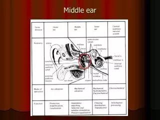
MIDDLE EAR CLEFT • Tympanic cavity • Eustachian tube • Mastoid antrum • Aditus • Mastoid air cells
Tympanic cavity • Irregular air filled space within the temporal bone • Contents • 3 ossicles: Malleus, incus and stapes • 2 muscles: Tensor tympani and stapedius • 2 nerves: Chorda tympani and tympanic plexus (IX) • Mucosal folds and ligaments • Vessels
Tympanic Cavity • EPITYMPANUM (ATTIC) • MESO TYMPANUM • HYPOTYMPANUM • Protympanum
Tympanic cavity may be thought of as a box with 4 walls, a roof and a floor
Roof of tympanic cavity • Tegmen Tympani - a thin plate of bone • Formed in part by petrous & part by squamous bone & Petro-squamous suture
Floor of Tympanic cavity • Thin plate of bone • Separates the tympanic cavity from the dome of the Jugular bulb • Structures passing – small opening for tympanic branch of Glossopharyngeal nerve
Lateral Wall • Part membranous & Part bony • Tympanic membrane forms the central portion • Above & below – bony outer lateral walls of epitympanum & hypotympanum
Medial Wall Separates the middle ear from the inner ear • Promontory – basal turn of cochlea • Oval window – opens into vestibule • Round Window – opens into scala tympani • Horizontal tympanic portion of facial nerve • Horizontal semicircular canal – above the facial nerve • Processuscochleariformis – Tendon of tensor tympani takes a turn here
Posterior wall • Opening to mastoid antrum • Pyramid • Facial recess (suprapyramidal recess) • Sinus tympani ( infrapyramidal recess)
Auditory ossicles • Conduction of sound waves from the external ear to middle ear • Links the tympanic membrane to oval window and cochlea
Malleus ( Hammer) • Largest of the three • Head • Neck • Anterior process – ant malleolar ligament • Lateral Process – receives anterior & posterior malleolar folds • Handle – between fibrous & mucosal layers of TM
Incus (anvil) • Has a body & 2 processes • Body- epitympanum • Short process lies in fossa incudis • Long process has a small medially directed lenticular process which articulates with the stapes
Stapes (stirrup) • Consists of a head, neck & crura • Head articulates with incus • Stapediusisertion into posterior part of neck and posterior crura • The 2 crura join the footplate
Muscles • Tensor tympani 1st arch muscle Supplied by mandibular nerve Attached to neck of malleus, tenses TM • Stapedius Supplied by facial nerve Attached to neck of stapes. Dampens loud sounds
Nerves • Chorda tympani • Tympanic plexus • Formed by tympanic branch of glossopharyngeal nerve and caroticotympanic nerves • Supplies medial surface of TM, tympanic cavity, ET and mastoid air cells • Also carries secrotomotorfibres to parotid