Download
1 / 19
300 likes | 852 Vues
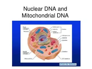
Nuclear DNA and Mitochondrial DNA. Purpose. Isolate, amplify, and sequence a piece of DNA from the mitochondria of your own cheek cells. Nuclear DNA. Present in almost every cell Combination from both parents; 23 chromosomes from each parent. Mitochondrial DNA.

E N D
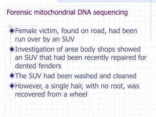
Purpose • Isolate, amplify, and sequence a piece of DNA from the mitochondria of your own cheek cells.
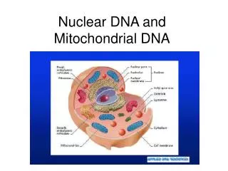
Nuclear DNA • Present in almost every cell • Combination from both parents; 23 chromosomes from each parent
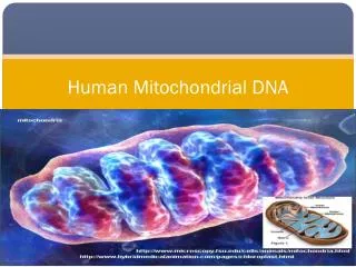
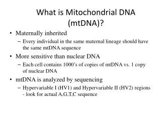
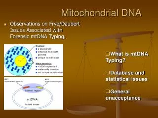
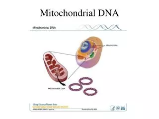
Mitochondrial DNA • Each cell contains thousands of mt, each containing copies of its DNA • Mt DNA is in larger quantities in a cell • Nuclear DNA is larger in size
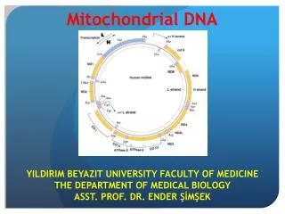
Mt DNA is 16,569 bases in length and consists of 2 different regions • Coding Region • Produces 13 proteins, 22tRNAs, 2rRNAs needed for cell respiration • This region has very little variability • So everyone’s DNA in this region will be nearly the same sequence of TGCAs
Control Region This region is highly variable within the human population Consists of 2 subregions HV1 = 342 bp HV2 = 268 610 bp with a lot of variability
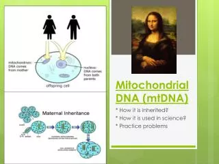
Mt DNA is inherited from mom • Every sibling will get their mt DNA from their mother • Why?
Why Mom? • Egg contains 23 chromosomes and cell cytoplasm which contains thousands of maternal mt • Sperm contains 23 chromosomes with very little cytoplasm
Zygote = Fertilized Egg • When egg and sperm join only female mt survive and are passed onto to new baby.
Mutations occur in the control region of mt DNA at a regular rate and are passed onto children by the mom.
How do we use this information? • We can compare DNA from the controlling region to other living humans • See how related to you are to each other • Compare to prehistoric remains of human fossils • Identify where you DNA originated • Identify ancestral relationships between modern populations • Compare your highly variable regions to other species
Mitochondrial Eve • Oldest women who would have donated her mtDNA to every ancestor in the world • Comparisons can be made by how many variations exist between her DNA and your DNA.
How is mtDNA isolated? • Isolate DNA from cheek cells • Complete a PCR reaction • Produce millions of extra copies of HV1 on the control region of mtDNA • Send amplified DNA away to be sequenced (Identify the exact sequence of TGCAs in HV1 in your mtDNA) • Compare your sequence ot classmates and database of prehistoric DNA
Procedure • Pour 10 ml of sports drink into mouth and vigorously swish for 1 minute. • Expel saline solution into a paper cup. • Swirl to mix cells in the cup and transfer 1 ml (1000 µl) of the liquid to 1.5 ml tube. • Place your sample tube, in a microcentrifuge, and spin for 1.5 minutes. • Carefully pour off supernatant into paper cup or sink. Be careful not to disturb the cell pellet at the bottom of the test tube. A small amount of saline will remain in the tube.
Resuspend cells in remaining saline by pipetting in and out. • Withdraw 30 µl of cell suspension, and add to tube containing 100 µl of Chelex. Vortex to mix. • Place tube in a thermal cycler for 10 minutes at 99°C. • After boiling, vortex tube. Place in a microcentrifuge and spin for 30 sec. • Transfer 30 µl of supernatant (containing the DNA) to clean 1.5 ml tube. Avoid cell debris and Chelex beads. • Store your sample on ice or in the freezer until ready to begin Step 12.
Use a micropipet with a fresh tip to add 22.5 µl of the appropriate primer/loading buffer mix to a PCR tube containing a Ready-To-Go PCR Bead. • Use fresh tip to add 2.5 µl of human DNA (from Part I) to reaction tube. Pipet up and down to mix the contents of the tube. • Label the cap of your tube with a number, as assigned by your teacher. • Store all samples on ice until ready to amplify according to the following profile:Denaturing time and temperature 30 sec - 94°C Annealing time and temperature 30 sec - 58°CExtending time and temperature 30 sec - 72°C Hold at -4°C (optional)
Use a micropipet with a fresh tip to add 15µl PCR sample/loading dye mixture into your assigned well of a 1% agarose gel. • Load 5 µl of the pBR322-BstNI size markers into one lane of gel. • Electrophorese at 130 volts for 20-30 minutes. Adequate separation will have occurred when the cresol red dye front has moved at least 50 mm from the wells. • Gel may be stained with 1 µg/ml ethidium bromide for 10 minutes followed by 20-30 minutes destaining with water.