Download
1 / 32
320 likes | 430 Vues
Explore the study of histology in Biology 523 with Dr. Elizabeth Connor. Learn about tissue identification, microscopy, and the relationship between structure and function. Join us in discovering the fascinating world of anatomical sciences!

E N D
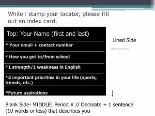
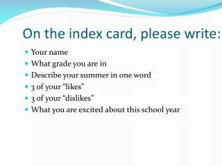
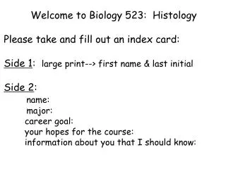
Welcome to Biology 523: Histology Please take and fill out an index card: Side 1: large print--> first name & last initial Side 2: name: major: career goal: your hopes for the course: information about you that I should know:
Biology 523: Histology, 4 credits Instructor: Dr. Elizabeth Connor 353 Morrill IV South telephone: 545-4855 email: econnor@bio.umass.edu office hours: by appointment
ANNOUNCEMENTS • Flu can include: fever, cough, sore throat, runny or stuffy nose, • body aches, headache, chills, and fatigue • Preventing flu• Get vaccinated. • Wash your hands often.• Cough or sneeze into a tissue or your elbow instead of into your hands. • Avoid touching your eyes, nose or mouth, to reduce the spread of germs.• Don't share food, drinks, utensils and other similar items.• If you’re sick, stay home!!! Email me about your situation and we will work it out.
OBJECTIVES #1- ANATOMY: To understand the structural basis for the functioning of a living organism Close relationship between structure and function
GROSS ANATOMY (macroscopic) HISTOLOGY (microscopic) ANATOMY
OBJECTIVES #2- TECHNICAL ADVANCES: To appreciate that our ability to answer questions is driven by the development of new technology
Why study histology? What are the uses of histological techniques and microscopic tissue identification?
Requirements: Tissue Preparation Start with an organism: Organ->Tissues-> Cells-> Molecules fixation/sectioning/ staining Imaging (Microscopy) Interpretation - cell biology - physiology - structure of tissues - organ structure/function Visual Integration **A challenging skill**
Teaching Assistants: Bryan Olson bolson@nsm.umass.edu Undergraduate Lab Assistants
Required Textbook: Histology: A Text and Atlas, Ross, Kaye, and Pawlina 6th Edition, Lippincott Williams & Wilkins ISBN 0-683-30242-6 *** BRING TEXTBOOK TO LAB ***
Many free online histology materials are available! Jejenum Links on Course Webpage Recommend Histology World Recommend MedPics UCSD
Course Web Page • https://bcrc.bio.umass.edu/courses/spring2012/biol/biol523/ • Class Preparation Page: Announcements • Reading Assignments • Syllabus • Class Notes: pdfs and ppt of class presentations • Laboratory: syllabus, lab exercises, project guides, • online tutorials, useful links • Review Materials • - Review questions • - Sample Exams and Answers • - Animations and useful links
Grading: 300 total possible points Two 1 hour in class exams (55 points each) 110 Final Exam 60 Homework Problems 10 Laboratory Attendance 15 Three Laboratory Practical Exams 55 ( 15, 15, 25 ) Contributions to Images2012 Gallery 20 Group Oral Project Presentation 10 Individual Written Project Report (2 parts) 20
Laboratory Experience (120 Points) Exercises using Prepared Slides Assessment: Three Practical Exams (15, 15, 25 points) Group Lab Project #1 As a class, we will assemble Images2012, an on-line image gallery of tissues, cells and organs examined during the course. (20 points) >2500 images from previous class efforts also available on our course website Assessment: Evaluation of postings to Images2012 (20 pts) Accuracy, labeling, quality of image
Group Lab Project #1 Images2012 Gallery: Your work is protected by- “Please Note: The imagery created by students in this course is licensed under the Creative Commons Attribution- NonCommercial - ShareAlike 3.0 Unported License. To view a copy of this license, visit http://creativecommons.org/licenses/by-nc-sa/3.0/ or send a letter to Creative Commons, 444 Castro Street, Suite 900, Mountain View, California, 94041, USA.”
Laboratory Experience (120 Points) Group Lab Project #2 Sectioning and staining of frozen and paraffin-embedded tissue of a rat organ. Techniques: microtome sectioning, frozen sectioning with cryostat, staining, immunohistochemistry, fluorescence microscopy, bright field microscopy, imaging, image processing. Assessment: Group Oral Presentation: In class (10 pts) Individual Written Project Report: (20 pts)
First lab: Tuesday, January 31st or Wednesday, February 1st To prepare for lab- Read Lab 1: Microscopy and Imaging Think about project partners (3/group)
Secrets of Success • -Attend every class and every lab. • -Come to class and lab prepared. • Ask questions. • Complete all assignments. • Review & study material after each lecture. • Answer review questions. • -Attend help and review sessions. • -Identify study partners.
TIMETIMETIMETIMETIMETIMETIMETIMETIMETIMETIMETIMETIMETIMETIMETIMETIMETIMETIMETITIMETIMETIMETIMETIMETIMETIMETIMETIMETIMETIMETIMETIMETIMETIMETIMETIMETIMETIMETI
ACCESS TO ISB 264 Exceptions: it will be in use by other courses on: Monday and Thursday 1:25pm-4:25pm Friday 3:35pm-4:35pm ……….And some other times to come
Challenge: Identify tissues. (Epithelial, Nervous, Muscle, Connective Tissue) Identify composite of tissues as an organ. Use appropriate descriptive language and terms. Understand the appropriate use of techniques to study material at the histological level
CROSS SECTION Orientation and Plane of section LONGITUDINAL SECTION
Challenges of Interpreting Plane of Section = Organ = Surrounding Tissues
Challenges of Interpreting Plane of Section = Organ = Surrounding Tissues