Patellar dislocation in adolescents
Patellar dislocation in adolescents. Mr Michalis Zenios Consultant Paediatric Orthopaedic Surgeon MBChB ( Hons ), MRCS (Eng), MSc, FRCS ( Orth ). Paediatric Orthopaedics. Fellow Sydney 2006-2007. Consultant Manchester 2007 -2012. Aims. Aetiology of patellar instability/ subluxation

Patellar dislocation in adolescents
E N D
Presentation Transcript
Patellar dislocation in adolescents Mr MichalisZenios Consultant Paediatric Orthopaedic Surgeon MBChB (Hons), MRCS (Eng), MSc, FRCS (Orth)
Paediatric Orthopaedics Fellow Sydney 2006-2007 Consultant Manchester 2007 -2012
Aims • Aetiology of patellar instability/subluxation • Assessment • Treatment (Evidence based) • Congenital patellar dislocation
Patellofemoral Instability Patellofemoral pain Patellofemoral subluxation Patellofemoral Dislocation
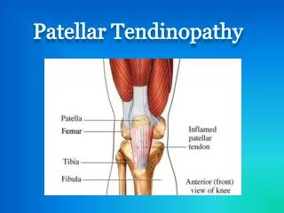
Patellofemoral Instability Bony Causes (local) • Femoral Trochlea • Patella Shape • Patella Height
Patellofemoral Instability Bony Causes (Lower Limb) • Genu Valgum • Femoral Torsion • Tibial Torsion
Patellofemoral Instability Soft tissue restraints • Medial • Medial patellofemoral (60%) • Medial Retinaculum • VMO • Lateral • Vastus Lateralis • Lateral Retinaculum
Pathology • Lateral hypermobility of the patella • Dysplastic distal one third of VMO • High or lateral position of the patella • Previous history of patellar subluxation
Patellar dislocations • Rare in a child. Common in adolescents. • Twisting injury or direct trauma • Lateral • Acute vs recurrent • Osteochondral fractures of patella or femur
Patellofemoral Instability Assessment • History • Acute or spontaneous • Duration • Number of episodes • Circumstances of injury • Previous treatment • Beware ACL injury (Pop) • Syndromes
Patellofemoral Instability Assessment • Examination • Full knee examination • Patella Tracking • J-sign • Medial or lateral tenderness • Tilt or lateral tightness • Apprehension Test (most reliable) • Q-angle • Torsional profile • General Laxity
Patellofemoral Instability Investigation • Plain X Rays • AP (? Osteochondral lesion) • Lateral view • 30 deg flexion (Koshino Index) • Merchant View • 30 deg flexion
Radiology • Insall index • < than o.8 suggests patella alta
Patellofemoral Instability • Sulcus Angle 140 degrees • Congruence angle -6 +/- 11degrees
Patellofemoral Instability • CT Scans • Fulkerson views • Vary knee flexion • MRI Scans • Medial restraints • EUA & Arthroscopy • Acute (MPFL) • Check tracking
Radiological measurements • Tibial tubercle trochlear groove distance • Lateralisation of the patella • Abnormal when above 20 mm
Patellofemoral Instability Conservative Treatment: • RICE • SLR/ Isometric Quadriceps • Open and closed chain kinetic exercises • Gradual return to activities • No casts or immobilization • Patellar stabilizing orthosis • Time
Patellofemoral Instability ? Role for acute surgery Treatment: • No place for acute operative stabilization in children and adolescents Acute patellar dislocation in children and adolescents. Surgical technique. J Bone Joint Surg Am. 2009 ; 91: 139-45. Nietosvaara Y, Paukku R, Palmu S, Donell ST. “The slaying of a beautiful hypothesis by an ugly fact” – T H Huxley
Acute patellar dislocation in children and adolescents: a RCT. J Bone Joint Surg (Am) 2008;90(3):463-470 62 patients younger than 16 who sustained acute patellar dislocation with an osteochondral fragment of 15mm. 36 0peratively 28 non-operatively: 1. 7 only lateral release 2. 29 repair medial structures
Acute patellar dislocation in children and adolescents: a RCT. J Bone Joint Surg (Am) 2008;90(3):463-470 • 14 year follow up • Initial operative repair did not improve the long-term outcome. 70 % re-dislocation rates • Positive family history was a significant risk factor for recurrence
Acute patellar dilsocation in adlescents: operative versus non-operative treatment. Int orthopaedics. Apostolovic 2011;35(10):1483-1487. • Non randomised prospective study- 37 adolescent knees • Decision for surgery on the basis of clinical and arthroscopic findings. Not clear • No difference between operative and non-operative treatment in terms of re-dislocation rates and functional outcome
Surgical intervention • Recurrent instability with functional compromise • Osteochondral lesions. Repair if > 2cm
Patellofemoral Instability • Surgical Strategy (100 operations in 100 years!) • Proximal Re-alignment (TUBS) • Acute initial episode • Lax soft-tissue restraints • Restore anatomy (MPFL reconstruction/ Insall procedure) • Distal Re-alignment (AMBRI) • Predisposition to patellar subluxation • Anatomical factors (Increased Q Angle) • Reconstruct anatomy • Patellar tendon or Tibial Tubercle
Patellofemoral Instability • Surgical Strategy (100 operations in 100 years!) • Proximal Re-alignment (TUBS) • Acute initial episode • Lax soft-tissue restraints • Restore anatomy (MPFL reconstruction/ Insall procedure) • Distal Re-alignment (AMBRI) • Predisposition to patellar subluxation • Anatomical factors (Increased Q Angle) • Reconstruct anatomy • Patellar tendon or Tibial Tubercle
A Surgical algorithm for the treatment of patellar dislocation. Results of 5 year follow up. ActaOrthopBelgica 2013.
A Surgical algorithm for the treatment of patellar dislocation. Results of 5 year follow up. ActaOrthopBelgica 2013. • Higher re-dislocation rates in immature patients who underwent proximal re-alignment procedures. • Mature patients with combined proximal and distal procedures had the lowest re-dislocation rates but low functional scores.
Predictors of recurrent instability after acute patellofemoral dislocation in paediatric and adolescent patients. Am J Sports Med 2013;41(3):575-581. USA. • 222 knees • Mean age 14.9 years • Patients with open physes and dysplastic trochlea had the highest dislocation rate at 69% • Age, sex, body mass index and patella alta were not associated with recurrent instability
Outcomes after patellar re-alignment surgery for recurrent patellar instability dislocations: a minimum 3-year follow-up study of children and adolescents. JPO 2011;31(1):65-71. USA • Recurrent dislocation 7% • Subjective opinion of knee function was less than expected 5 years post-op.
Weight-bearing osteochondral lesions of the lateral femoral condyle following patellar dislocation in adolescents athletes. Orthopaedics 2012;35(7):1033-1037. USA • 80 patients with acute patellar dislocation • 27.5% had an osteochondral lesion of the wt bearing area of lateral femoral condyle and 60% required surgical intervention • Suggestion of performing an MRI if there is tenderness over the lateral femoral condyle.
Surgical treatment for instability -Summary • Do not operate acutely • Understand and try to correct your anatomy • No tibial tubercle transfer in skeletally immature patients
Congenital patella dislocation • First described by Singer 1856 • Present at birth diagnosed then or within first decade • The patella should be permanently fixed to the lateral aspect of the femur
Congenital patella dislocation Aetiology Failure of the myotome containing the Quadriceps and Patella from internally rotating in the first trimester
Congenital patella dislocation Pathology • Extensor mechanism inserted antero-laterally • Contracture of Iliotibial band, Vastuslateralis, and Lateral capsule • Loose and atrophic medial capsule & VMO • Hypoplastic femoral trochlea • External rotation of tibia and valgus deformity of knee
Congenital patella dislocation Treatment • Initiated before 1st birthday • Extensive lateral release of whole of Vastus lateralis & knee capsule • Extensor mechanism is reduced and medial structures lateralised +/- Roux Goldthwaite