Surgical dislocation
Surgical dislocation. Vasu Pai [ VOL. 83-B, NO. 8, NOVEMBER 2001].

Surgical dislocation
E N D
Presentation Transcript
Surgical dislocation Vasu Pai [VOL. 83-B, NO. 8, NOVEMBER 2001]
Diagram showing the line of trochanteric osteotomy for the trochantericflip. Proximally, the osteotomy exits just anterior to the most posteriorinsertion of gluteus medius. Distally, the entire origin of vastus lateralisremains on the trochanteric fragment (GMED, gluteus medius; PI, piriformis;OI, obturator internus; Q, quadratus femoris; VLAT, vastuslateralis).
Diagram showing that in slight flexion and external rotation of the femur(arrow) the trochanteric fragment, including the tendon of gluteus minimus,is flipped over anteriorly. The interval between gluteus minimus andthe tendon of piriformis is then developed and gluteus minimus retractedsuperiorly to expose the capsule (GMIN, gluteus minimus; C, capsule;GMED, gluteus medius; PI, piriformis; OI, obturator internus).
Operative technique In the lateral decubitus position, a lateral approach The fascia lata split accordingly. The leg is then internally rotated and the posterior border of gluteus medius identified. No attempt is made to mobilise gluteus medius or to visualise the tendon of piriformis. An incision is made from the posterosuperior edge of the greater trochanter extending distally to the • posterior border of the ridge of vastus lateralis. A trochanteric osteotomy with a maximal thickness of about 1.5 cm is made along this line with an oscillating saw. At its proximal limit, the osteotomy should exit just anterior to the most posterior insertion of gluteus medius. This preserves and protects the profundus branch of the MFCA, which becomes intracapsular at the level of the superior gamellusmuscle.
The greater trochanteric fragment is mobilised anteriorly with its attached vastus lateralis The most posterior fibres of gluteus medius are also released from the remaining trochanteric base. The osteotomy is correct when only part of the fibres of the tendon of piriformis has to be released from the trochanter With the leg flexed and externally rotated, vastus lateralis and intermedius are elevated The inferior border of gluteus minimus is separated from the relaxed piriformis and underlying capsule. The constant anastomosis along the distal border of the piriformis muscle and tendon, is preserved. Care has to be taken to avoid injury to the sciatic nerve. The entire flap, including gluteus minimus, is retracted anteriorly and superiorly to expose the superior capsule. This is facilitated by further flexion and external rotation of the hip. The anterior, superior and posterosuperior capsule can now be visualised .
Diagram showing that for the Z-shaped capsulotomy the femur is flexedand externally rotated further (arrows). All external rotators are leftintact.
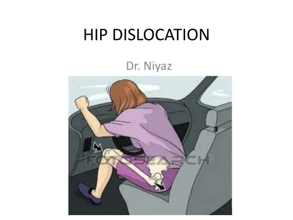
Diagram showing that for subluxation and dislocation of the femoral headthe hip is flexed, externally rotated and the leg brought over the front ofthe operating table and placed in a sterile bag
The anterior dislocation is completed after the ligamentum teres is either torn by external rotation, or incised. The stump of the ligament remaining on the femoral head may be resected. The foveolar artery, which is frequently patent in the ligamentum teres, is not an important source By manipulating the leg, the surgeon now has 360° access to the acetabulum and head For a complete inspection of the acetabulum three retractors are used (Fig. 5). The knee is elevated with an assistant applying axial pressure to bring the femoral head posterior to the acetabulum. No retractors are needed for visualisation of the femoral head, the knee being merely lowered to allow the head to rise out of the surgical wound. For its most posterior aspect a blunt Hohmann retractor around the neck may be useful. The retinaculum protecting the terminal branches of the MFCA to the femoral head is clearly visible on the posterosuperior aspect of the neck as a mobile layer of connective tissue.
The labrum is inspected and probed, and the articular surfaces of the femoral head and acetabulum examined. Bleeding of the surfaces of the cancellous bone after trimming osteophytes on the periphery of the head are further signs of satisfactory vascularity. During the exposure the articular cartilage is constantly irrigated with Ringer lactate solution Reduction of the hip may easily be accompanied by manual traction on the flexed knee and internal rotation. The capsule of the hip can be repaired, but not tightened since this may create tension on the retinacular vessels leading to a drop in the perfusion The greater trochanter is reattached using two or three 3.5 mm cortical screws or cerclage wire. When an intertrochanteric osteotomy is undertaken the trochanteric fragment is transfixed by the
Inspection of the acetabulum one retractor is impacted above the acetabulum. One retractor hooks on the anterior rim and a third retractor levers the calcar of the neck against the incisura acetabuli. For inspection of the femoral head no retractorsare needed, the knee is lowered and with rotation of the leg (arrows) different surfaces of the head can be visualised.
Complications • 1.A partial neurapraxia of the sciatic nerve was diagnosed after operation; both resolved within six months without residual sequelae. • 2.Trochanteric fixation failed in three patients, requiring a second operation. • 3. Heterotopic ossification was seen in 37%.
Using an anterior (Smith-Petersen) approach the femoral head can be dislocated safely, but inspection of the acetabulum is limited, unless the tensor fascia lata and gluteus medius are extensively detached from their origins. • Anterolateral and direct lateral approaches may allow dislocation of the femoral head, but again exposure of the acetabulum is difficult and incomplete. • With the posterior approach, tenomyotomyof the external rotator muscles is necessary, which interrupts the anastomosis between the inferior gluteal artery and the deep branch of the MFCA. • Trochanteric osteotomy requires more care in regard to union, since there is no balancing of the force of gluteus medius by vastus lateralis; the myofascialflap approach needs special attention until the resuturedsoft tissues have healed.