Download

1 / 100
2.26k likes | 7.23k Vues
DISORDERS OF THE GASTROINTESTINAL SYSTEM. DR ADIBE MAXWELL CLINICAL PHARMACY 2017. Anatomy & Physiology of the Gastrointestinal System. GI tract: breakdown, absorption and elimination Upper potion the mouth esophagus stomach Lower portion small intestine

E N D
DISORDERS OF THE GASTROINTESTINAL SYSTEM DR ADIBE MAXWELL CLINICAL PHARMACY 2017
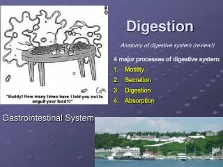
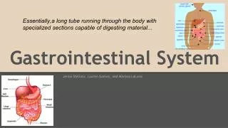
Anatomy & Physiology of the Gastrointestinal System • GI tract: breakdown, absorption • and elimination Upper potion • the mouth • esophagus • stomach Lower portion • small intestine • large intestine • rectum • anus. (Day et al., 2010)
DIGESTIVE SYSTEM • FUNCTIONS: ingest food • DIGESTION:break it down into small molecules • ABSORPTION:absorb nutrient molecules • ELIMINATION:eliminate nondigested wastes • ASSESSORY ORGANS : • pancreas, liver, gallbladder
Disorders of the upper GI systemDisorders affecting Ingestion • ANOREXIA: lack of appetite, could be from emotional or physical factors • lab tests may be done to assess nutritional status • Medical treatment: supplements may be ordered, TPN or enteral feedings Interventions: • oral hygiene, clean room, determine cause of nausea and treat, include family and friends(socialization), respect likes and dislikes, education
STOMATITIS • Inflammation of the oral mucosa (mouth) • Causes: trauma, organisms, irritants, nutritional deficiency, diseases, chemotherapy • S/S: swelling, pain, ulcerations, excessive salivation, halitosis, sore mouth • Treatment: • pain relief, removal of causative factor, oral hygiene, medications, soft bland diet
GINGIVITIS • Inflammation of the gums • Causes: poor oral hygiene, poorly fitting dentures, nutritional deficiency • S/S: red, swollen, bleeding gums, painful • Treatment: dental hygiene, prevention of complications
Interventions:Stomatitis and Gingivitis • Assess mouth condition • Administer medications • Mouth care • Soft bland diet, no spicy foods • Observe for complications • Teach importance of mouth and gum care
HERPES SIMPLEX TYPE 1 • Infection affecting the lips and mucous membranes of the mouth • Causes: Herpes simplex virus • S/S: Vesicles on the mouth, nose or lips, malaise, edema of surrounding area • Treatment: Antiviral medication(Zovirax), analgesics, symptomatic relief • Interventions: Administer meds, keep lesions dry, provide symptomatic relief
LEUKOPLAKIA • Abnormal thickening and whitening of the epithelium of the mucous membranes of the cheeks and tongue • Causes: Chronic irritation • S/S: Thickened white or reddish lesions on the mucous membrane, lesions can not be rubbed off
Treatment: May be surgically removed or treated with chemotherapy, meticulous oral hygiene • Interventions: Assess mouth frequently, assist with oral hygiene, discuss removal of sources of irritation
ORAL CANCER • Malignant lesions may develop on the lips, oral cavity, tongue and pharynx. Generally squamous cell carcinomas • Causes: high alcohol consumption, tobacco use, external irritants • S/S: Leukoplakia, swelling, edema, numbness, pain • Diagnosis: biopsy
Treatment: • Surgery • Radiation or chemotherapy • depends on the size and location and the lesion • Interventions: consult MD for special mouth care, monitor respiratory status, administer pain med, assess ability to swallow and talk, assess for infection at incision site, education
ESOPHAGITIS • Inflammation or irritation of the esophagus • Causes: Reflux of stomach contents, irritants, fungal infections, trauma, malignancy, intubation • S/S: heartburn, pain, dysphagia • Treatment: treat underlying cause • Interventions: soft bland diet, administer meds, observe for complications
ESOPHAGEAL VARICIES • Tortuous, distended vessels of the esophagus • may rupture and bleed • causes: Portal hypertension caused by cirrhosis of the liver • S/S Hematemesis, hemorrhage from UGI, black tarry stools, pain, shock
Treatment: • Iced saline lavage • Medications( Vasopressin, antibiotics, analgesics) • Surgeries: ligation, injection sclerotherapy • Blood transfusions
Interventions: • administer meds • provide pre/post op care • administer blood transfusions • monitor tube placement • assess vital signs, bleeding
CANCER OF THE ESOPHAGUS • Prognosis is very poor, diagnosed at late stages • Causes- no known cause, predisposing factors; irritation, poor oral hygiene • S/S- progressive dysphagia, painful swallowing, weight loss, vomiting, hoarseness, coughing, iron deficiency, anemia, occult bleeding or hemmorage
Treatment of CA of Esophagus • Palliative treatment is common • Radiation, chemotherapy • surgery: • Esophagectomy • Esophagogastrostomy • Esophagoenterostomy • Gastrostomy
Interventions • Maintain NG tube after surgery • Assess for signs of hemorrahage • Monitor respiratory status • monitor adequacy of nutritional intake ( high protein, high calorie diet) • assess ability to swallow • allow patient to ventilate feelings
DISORDERS OF DIGESTION AND ABSORPTION • N/V • Hiatal Hernia • Gastritis • Peptic Ulcer • Stomach Cancer
NAUSEA AND VOMITING • Nausea: unpleasant sensation usually preceding vomiting, may have abdominal pain, pallor, sweating, clammy skin • Causes: irritating food, infection, radiation, drugs, hormonal changes, surgery, inner ear disorders, distention of the GI tract
Vomiting: forceful expulsions of stomach contents through the mouth. Occurs when vomiting reflex in the brain is stimulated. • Projectile vomiting- is forceful ejection of stomach contents. • Regurgitation- gentle ejection of stomach contents without nausea or retching
Complications and Treatment • May lead to dehydration, metabolic alkalosis, aspiration • Treatment: Antiemetics( Phenergan, Dramamine, Scopolamine patch Reglan), IV fluids, NG tube, TPN • Intervention: through assessment, keep patient comfortable, offer liquids, position on side, suction setup in the room
HIATAL HERNIA • Protrusion of the lower esophagus and stomach upward through the diaphragm into the chest • SLIDING-gastroesophageal junction above the hiatus • ROLLING( paraesophageal)-junction in place portion of stomach rolls up through diaphram • Causes; weakness in the lower esophageal sphincter, related to increased abdominal pressure, long term bedrest, trauma
Signs and Symptoms • Feelings of fullness • dysphagia • eruption • regurgitation • heartburn • Complications: Ulcerations, bleeding, aspiration • seen in 50% of people over 60.
Treatment for Hiatal Hernia • Drug therapy • H2 receptor antagonists:Tagamet,Zantac, Pepsid- reduce stomach secretions • Antacids- neutralize stomach acids • Reglan, Propulsid- increase stomach emptying • diet therapy- decrease caffeine fatty foods, alcohol( reduce LES tone), acidic and spicy foods
SURGERY • Nissen Fundoplication • Angelclikprothesis CARE: • assessment, pain relief, watch for aspiration, nutrition, education
GASTRITIS • Inflammation of the lining of the stomach • ACUTE: excessive intake of food or alcohol. Food poisoning, chemical irritation • CHRONIC: repeated episodes of acute, H Pylori
Signs/Symptoms and Complications • Nausea, vomiting, feeling of fullness, pain in stomach, indigestion. With chronic may have only mild indigestion • changes in stomach lining with decrease in acid and intrinsic factor ( high risk for pernicious anemia)
Treatment • Treat symptoms, and fluid replacement • Medications: antacids, H2 receptor blockers, B 12 injections, corticosteroids analgesics, antibiotics if H Pylori • bland diet, frequent meals • Eliminate the cause • surgical intervention • BEST DIAGNOSIS IS GASTROSOPY & BIOPSY
CARE • Good HX and review of present S/S • pain relief, adequate nutrition, hydration, stress management, education
PEPTIC ULCER • Loss of tissue from the lining of the digestive tract. May be acute or chronic. • Classified as gastric or duodental (stress- develop 24-48hr. After event) • CAUSES: drugs, stress, heavy alcohol and tobacco use, infection (H .pylori bacteria) Conditions that cause high gastric acid concentration
Gastric Ulcers burning pain 1-2 hrs. after meals, upper left abd/back,relieved by food N/V, anorexia, wt loss Shallow/ gastric secretions deceased Older men, working class, bld type A, under stress Duodenal Ulcers burning/ cramping pain 2-4hrs. P meal, beneath xiphoid and back, relieved by antacids/food increased gastric acid Young men, all social classes, bld type O, chronic illnesses Peptic Ulcer comparison
PEPTIC ULCER COMPLICATIONS • HEMORRHAGE • PERFORATION • PYLORIC OBSTRUCTION
TREATMENT • Drug therapy • Antacids • H2 RECEPTOR BLOCKERS • ANTICHOLINERGICS-Pro-Banthine, Robinul, Bentyl • SUCRALFATE- Carafate • Antibiotics –Flagyl, tetracycline, Biaxin • treatment goals- relieve symptoms, promote healing, prevent complications and recurrence
Interventions • Three meals a day – decreases acid production • decrease foods that stimulate acid secretions and cause discomfort • treat pain with rest, diet and drug therapy • educate on stress management and relaxation
Surgical options for gastric ulcers • To decrease acid secretion: • vagotomy • pyloroplasty • gastroenterostomy • antrectomy • subtotal gastrectomy • Billroth I • Billroth II
care after gastric surgery • No signs of complications • Gastric dilation • Obstruction • Perforation • Maintenance of NG tube: • Suction • do not irrigate or reposition tube • type of drainage
Adequate nutrition: • NPO gradually advance from clear liquids to full liquids then solid foods • Assess for N/V, abdominal distention • Size of meals changes depending on type of surgery • Gastric surgeries can have serious effects on absorption of vit. B12, folic acid, iron, calcium, vit, D
education • Reinforce diet • teach signs of complicatons • Avoid risk factors
STOMACH CANCER • Rare (25,000/yr.), common in males, African American, over 70 and low socioeconomic status. 60% decrease in past 40 yrs. • No S/S in early stages • Late stages S/S: N/V, ascities, liver enlargement, abd. Mass • Mets to bone and lung • 10% survival rate after 5 yrs.
Risk factors: pernicious anemia, chronic gastritis, cigarette smoking, diet high in starch, salt, salted meat, pickled foods, nitrates • Treatment: surgery/ chemotherapy/ radiation • subtotal gastrectomy, total gastrectomy
Inflammatory Bowel Disease • IBD refers to two chronic inflammatory GI disorders: Crohn’s disease and Ulcerative Colitis, both cause inflammation and ulcerations of the intestine. • Two Types: • Crohn’s: usually affects the intestines. • Ulcerative Colitis: usually affects the large intestine. • Statistics: • 200,000 Canadians have IBD • 15-30 years of age at highest risk. • gender nonspecific. (Day et al., 2010)
Inflammatory Bowel Disease: Complications • Ulcers: chronic inflammation can lead to open sores within the digestive tract. • Fistulas: when an ulcer forms and extends completely through the intestinal wall. • Anal fissures: crack or cleft in the anus or skin where infection occurs. • Malnutrition: difficulties eating and absorbing nutrients. • Other problems: • Arthritis • Kidney and gallstones • Inflammation of eyes and skin (Day et al., 2010)
Crohn’s Disease: Etiology & Pathophysiology • Chronic disorder that causes inflammation of the GI tract, most commonly affecting the small intestine. • Transmural; affecting all layers of the mucosa. • Begins with edema and thickening of the mucosa. • Ulcers appear on the inflamed mucosa, causing fistulas and fissures. • Scaring, thickening and narrowing of the GI tract. • Statistics: • Usually diagnosed in adolescents • Prevalence has risen in the past 30 years. • seen more in smokers (MFMER, 2011 ; Day et al., 2010; CCRC, 2008; CCFC, 2008 ; Mahan & Escott-Stumop, 2004;)
Crohn’s Disease: Clinical Manifestations • Persistent diarrhea • Loss of appetite & weight loss • May have rectal bleeding • Cramping abdominal pain • Steatorrhea • Fatigue • Fever Complications • Bowel obstruction • Sores of ulcers • Fistulas • Malnutrition (CSIR, 2012; CCFC, 2008)
Crohn’s Disease: Diagnosis • Health history: Onset, associated symptoms, pain, stool, & rectal bleeding. • Blood tests: anemia or infection and certain antibodies • Fecal occult blood test: looking rectal bleeding. • Stool sample : presence of white blood cells. • Colonoscopy : visualize and collect biopsy. • Flexible sigmoidoscopy : examine sigmoid colon. • Barium enema : evaluate large intestine with x-ray. • X-ray : rule out toxic megacolon. • CT scan : assess for complications and amount of infection. • MRI : diagnosis and management. • Capsule endoscopy : all other diagnostics are negative. • Double – balloon endoscopy : still questioning diagnosis. • Small bowel imaging : locate narrowing or inflammation. (CSIR, 2012; MFMER, 2011; CCFC, 2008)
Colonoscopy A procedure used to see inside the colon and rectum • Used to investigate intestinal signs and symptoms. • Preparation: • Bowel prep to empty the bowel. • No solid food the day before • Laxative or enema kit • Adjust medications • Postoperative: • Hour to recover • Blood with first BM • When to seek medical care • Severe abdominal pain, fever, dizziness, weakness, bloody BM’S http://www.youtube.com/watch?v=rSXTIzqWc7s (NIDDK, 2011; MFMER, 2011)
Flexible Sigmoidoscopy A procedure used to evaluate the part of the large intestine and investigate signs and symptoms. • Preparation: • No solid foods • NPO after midnight • Laxative or enema kit • Adjust medications • What to expect: • Usually does not require sedation or pain medication. • May feel abdominal cramping or urge to push. • Ability to take biopsies . • Takes about 15 minutes. (NIDDK, 2011; MFMER, 2011)
A special X-ray used to detect changes or abnormalities in the large colon and part of the small intestine. • Single-column: allows visualization of silhouette , shape and condition of colon. • Air-contrast: Air expansion improves the quality of X-ray images. • During exam: • No sedation necessary. • Side lying position. • May manipulate the colon manually. • Enema tube is inserted with a barium bag. • After exam: • May expel additional barium and air with BM. • Drink plenty of fluids, laxative may be required. Barium Enema (NIDDK, 2011; MFMER, 2011)