Cell Review/Chapter 3
Cell Review/Chapter 3. By Becky . Cytology is the study of the structure and functions of cells. The cell was first discovered by Robert Hooke while viewing a dried cork. The cells have been researched for 175 years. This research has led to the cell theory. . The Cell.


Cell Review/Chapter 3
E N D
Presentation Transcript
Cell Review/Chapter 3 By Becky
Cytology is the study of the structure and functions of cells. • The cell was first discovered by Robert Hooke while viewing a dried cork. • The cells have been researched for 175 years. • This research has led to the cell theory. The Cell
The cell theory has five basic concepts. • 1. Cells are the building blocks of all plants and animals. • 2. Cells are produced by the division of preexisting cells. • 3. Cells are the smallest units that perform all vital physiological functions. • 4. Each cell maintains homeostasis at the cellular level. • 5. Homeostasis at the tissue, organ, system, and individual levels reflects the combined and coordinated actions of many cells. The Cell Theory
Outside the cell is a fluid known as Extracellular Fluid. This fluid is a watery medium where the cell floats. • The Cell Membrane separates the cell’s contents and cytoplasm from the extracellular fluid. • The cytoplasm can further be broken down into a fluid known as Cytosol. • The cytoplasm has a very different composition than the extracellular fluid and must be kept separate. Cell Fluid
The cytoplasm includes a lot more protein than the extracellular fluid. • The cytosol contains a high concentrations of potassium, while extracellular fluid contains a high concentration of sodium. • The cytosol contains dissolved nutrients, ions, soluble and insoluble proteins, and waste products. • Cytosol has about 200 amino acids while as the Extracellular fluid only has 30. • There are major differences between the cytoplasm and extracellular fluid which need them to be separated. Cytoplasm VS. Extracellular Fluid
The cell membrane ( also known as the Plasma Membrane) forms the outer boundary of the cell. It is the cell’s bodyguard. • The major components of the cell membrane are phospholipids, proteins, glycolipids, and cholesterol. • The cell membrane is also a Phospholipid bi-layer because the phospholipids form two distinct layers. • Ions and water soluble compounds cannot enter the interior of the membrane because the lipid tails of the phospholipid molecules are hydrophobic. The Cell Membrane
Peripheral proteins are attached to the inner membrane surface of the cell membrane. • Internal proteins are embedded in the membrane. • Some of the internal proteins form channels that let water molecules, small water- soluble compounds and ions into or out of the cell. • The cell membrane also contains a variety of receptors that allow the cell to recognize and respond the specific molecules in its environment. Cell Membrane
Cells work together to maintain homeostasis at the tissue, organ, and system levels. • The essential communication and coordination activities involve the cell membrane, which forms the interface between each cell and the cell’s surroundings. • Regulation is essential because the intercellular and extracellular environments are quite different. Those differences must be maintained to preserve homeostasis. The cell and its environment
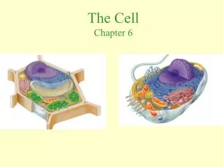
Organelles are found in most cells that are able to grow and reproduce. • Each organelle performs a function that is essential to normal cell structure, maintenance, and the metabolism. • Organelles can be divided into two categories: Nonmembranous organelles and membranous organelles. Organelle
Cytoskeleton Microfilament Are slender protein strands, commonly composed of the protein called actin. In most common cells, microfilaments are scattered throughout the cytoplasm. They form a dense layer under the cell membrane. Have two major functions: Anchor the cytoskeleton to integral proteins of the cell membrane. Actin microfilaments can interact with filaments composed of another protein called Myosin. • An internal protein framework that gives the cytoplasm strength and flexibility. • It has four major components: • Microfilaments, intermediate filaments, thick filaments, and microtubules. Nonmembranous organelles.
Intermediate Filaments Thick filaments Are relatively massive strands composed of myosin protein subunits. They are also abundant in muscle cells. This is where they react with actin filaments to produce powerful contractions within the cell. • Are defined chiefly by their size: their composition varies from one cell type to another. • They provide strength, stabilize the positions of organelles, and transport materials within the cytoplasm. • A specialized filament is called neurofilaments.
Microtubules Microvilli Are small, finger-shaped projections of the cell membrane. Are found in cells that are engaged in absorbing materials from the extracellular fluid. Are important because they increase the surface area exposed to the extracellular environment. • Are hollow tubes built from the globular protein called tublin. • They have a variety of functions: • Form the main components of the cytoskeleton, giving the cell strength and anchoring the organelles into the right position.
Centrioles Cilia contains nine pairs of microtubules surrounding a central pair. Cilia are anchored to a compact basal body located just beneath the cell’s surface. Cilia lining the respiratory tract beat in a synchronized manner to move sticky mucus and trapped dust particles towards the throat and away from the delicate respiratory surfaces. • Is a cylindrical structure composed of short microtubules. • All animal cells are capable of reproducing themselves contain a pair of centrioles. • The centrosome is the cytoplasm surrounding this pair. • Centrioles direct the movement of DNA strands through the cytoplasm.
Flagella Ribosomes Small, dense structures that cannot be see clearly with a light microscope. They are found in all cells. Consists of about 60% Rna and 40% protein. Two major types of ribosomes: Free Ribosomes Fixed Ribosomes • Resemble cilia but they are bigger than cilia • Moves a cell through the fluid surrounding the cell, rather than moving the fluid past the cell. • The sperm cell is the only human cell that has a flagellum.
Mitochondria Nucleus The control center for cellular operation. Most cells contain a single nucleus, but there are exceptions. A nuclear envelope surrounds the nucleus and separates it from the cytosol. The nuclear envelope is a double membrane containing a narrow pronuclear space. Chemical communication between the nucleus and cytosol occurs through the nuclear pores. • Small organelles that have an unusual double membrane. • The inner membrane contains many fold called Cristae. • Cristae increase the surface area exposed to the fluid contents (Matrix). • Respiratory enzymes are attached to the cristae and produce most of the ATP generated by mitochondria. Membranous Organelles
Endoplasmic Reticulum Golgi Apparatus Consists of flattened membrane discs, called saccules. Major Functions: Synthesis and packaging of secretions Packing of special enzyme Renewal or modification of the cell membrane. Materials move from saccule to saccule by means of small transfer vesicle. Vesicles containing secretions that will be discharged from the cell and are called secretory vesicles. The ejection process is called exocytosis. • Is a network of intracellular membranes. • Three major functions: • Synthesis: manufactures protein, carbs, and lipids • Storage: hold synthesized molecules • Transport: materials travel through place to place. • The ER forms hollow tubes, flattened sheets, and round chambers called Cisternae. • There are two types of ER: • Rough ER • Smooth ER
The cell has many way to obtain energy. • It can receive energy through lipids, carbohydrates, and proteins. • Lipids are fats and carbohydrates are your typical carbs that provide you with energy. They also provide the cells with the same energy so that their organelles can function correctly. • The organelle, Mitochondria, provides 95% of the energy needed to keep a cell alive. • It produces ATP through the breakdown of organic molecules in a series of reactions that also consume oxygen and generate carbon dioxide. Cell’s Energy
Lysosomes Peroxisomes Are smaller than lysosomes and carry a different group of enzymes. They absorb and neutralize toxins that are absorbed from the extracellular fluid or generated by chemical reactions in the cytoplasm. • Are vesicles filled with digestive enzymes. • They may function in the defense against disease. • This is called endocytosis. • When enzymes rapidly destroy the proteins and organelles of the cell it is called autolysis.
The nucleus is the control center for the cell. • The nucleus directs processes that take place in the cytosol and must in turn receive information about conditions and activities in the cytosol.. • In the nucleoplasm in the nucleus is where the DNA and RNA are found. • Our cell nuclei contains 23 chromosomes which is what makes a person look the way they do. • The nucleus controls the actions of the organelles within the cell. • The nucleus copies the chromosomes when it is time for the cell to divide. The Nucleus
The lifespan of a cell varies from hours to decades. • This depends on the type of cell and the environmental stresses involved. • A typical cell does not live near as long as a typical person, so over time cell populations must be maintained by cell division. • Even when development has been complete cell division continues to be essential to survive. Cell Life Cycle
There are two types of cell division. • Central of cell reproduction is the accurate duplication of the cell’s genetic material and its distribution to the two new daughter cells formed by division. This is called Mitosis. • Mitosis occurs during the division of somatic cells. • Somatic cells include all the cells in the body other than the reproductive cells, which give rise to sperm or eggs. • Most cells spend only a small part of their time actively engaged in cell division. • Somatic cells spend the majority of their functional lives in interphase. Cell Division
During interphase the cell performs all of its normal functions and making preparations for division. • There are numerous stages during interphase: • G0 phase • G1 phase • S phase • G2 phase • Gm phase • An interphase cell in the G0 Phase is not preparing for mitosis. Interphase
A cell that is going to divide first enters G1 Phase. • In this phase, the cell produces enough mitochondria, centrioles, cytoskeleton elements, endoplasmic reticulum, ribosomes, golgi membranes, and cytosol to make two functional cells. • In cell dividing at top speed, G1 may last as little as 8-12 hours. • When preparations have been completed, the cell enters the S phase. • Over the next 6-8 house the cell duplicates its chromosomes. Interphase
Throughout the life of a cell, the DNA strands in the nucleus remain intact. DNA synthesis or DNA Replication occurs in cells preparing to undergo mitosis or meiosis. • The goal of replication is to copy the genetic information in the nucleus so that one set of chromosomes can be given to each of the two daughter cells produced. • Once DNA replication has been completed, there is a brief 2-5 hours G2 phase devoted to last minute protein synthesis. • The cell then enters the Gm phase and mitosis begins.
Stage 1: prophase begins when the chromosomes of the cell coil so tightly that they become visible. • As a result of DNA replication during the S phase, there are two copies of each chromosome called chromatids. • They are connected at a single point called the centromere. • Spindle fibers begin to form between the centriole pairs. • Prophase ends with the disappearance of the nuclear envelope. Mitosis
Stage 2: Metaphase. The chromatids now move to a narrow central zone called the metaphase plate. • A microtubule of the spindle apparatus attaches to each centromere. • Stage 3: Anaphase. As if responding to a single command, the chromatid pairs separate and the daughter chromosomes move towards opposite ends of the cell • Stage 4: Telophase. This stage is in many ways the reverse of prophase. • The nuclear membranes form, the nuclei enlarge, and the chromosomes gradually uncoil.
Once the chromosomes disappear, nucleoli reappear and the nuclei resemble those of interphase cells. • Telophase marks the end of mitosis, but the daughter cells have yet to complete their physical separation. • This separation process is called cytokinesis. • As the daughter chromosomes near the end of the spindle apparatus, the cytoplasm constricts along the metaphase plate. • This process continues through Telophase and the completion of cytokinesis marks the end of cell division. • Meiosis is the cell division of sex cells. • It has the same phases, but creates four daughter cells instead of two. • They reproduce sex cells instead of somatic cells.
Two factors, one passive and one active, interact to create and maintain the Transmembrane potential. • The Transmembrane potential is a characteristic of all living cells because it results from the active and passive properties of their cell membranes. • The Transmembrane potential is just as important as any structural characteristics or organelles. • Many cell functions that involve the cell membrane involve changes in the Transmembrane potential. • But the Transmembrane potential has its own significant functions. Transmembrane Potential
In addition to the open sodium and potassium channels it has, the cell membrane contains gated ion channels that are closed at the normal resting potential. • Because the concentration gradients are large, any stimulus that opens one of these gates will produce a sudden rush of ions into or out of the cell. • If it can affect a channel protein, even a relatively weak stimulus can have a significant impact on the cell. • Because the Transmembrane potential can increase a stimulus in this way, it greatly increases the cell’s sensitivity to its environment. • The Transmembrane is maintained by the sodium-potassium exchange pump in the membrane. It makes sure the sodium and the potassium in the Transmembrane potential is stabilized.
Most cells in the body are firmly attached to other cells or to extracellular protein fibers. The attachment occurs at cell junctions that are not involved in membrane flow. There are four types of cell junctions: gap junctions, tight junctions, intermediate junctions, and desmosomes. • Gap junction: in a gap junction the two cells are held together by an interlocking of membrane proteins. • Gap junctions are most common in cardiac muscle ad smooth muscle tissue. They are also occasionally found between nerve cells • Tight junctions: at a tight junction, there is a partial fusion of the lipid portions of the two cell membrane. Attachment of Cells
Because the membrane is fused together, tight junctions are the strongest intercellular connections. • The tight junction also blocks the passage of water or solutes between cells. • Intermediate junctions: at an intermediate junction the opposing cell membranes, while remaining distinct, are held together by a thick layer of proteoglycans. • The cytoplasm at an intermediate junction contains a dense network of microfilaments that anchor the junction to the cytoskeleton. • This arrangement adds strength and helps to stabilize the shape of the cell.
Desmosomes: at desmosomes there is a very thing proteoglycan layer between the opposing cell membranes, reinforced by a network of intermediate filaments that lock the two cells together. • Desmosomes are very strong and the connection can resist stretching and twisting. • The desmosomes create links so strong that even dead skin cells are usually shed in thick sheets rather than individually. • Junctional complexes: cells lining the digestive tract, respiratory tract, or other passageways are held together by Junctional complexes. A single Junctional complex consists of a tight junction, an intermediate junction, and a desmosome, with the tight junction closest to the surface.