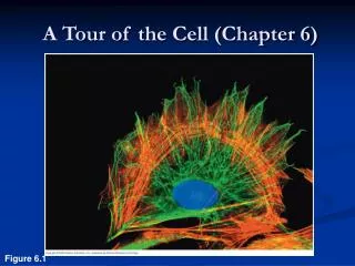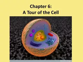
The Cell Chapter 6
1.1k likes | 1.31k Vues
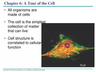
The Cell Chapter 6. How do we know about cells?. Microscopes: windows to the world of the cell. The discovery and early study of cells progressed with the invention and improvement of microscopes in the 17th century.

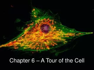
The Cell Chapter 6
E N D
Presentation Transcript
Microscopes: windows to the world of the cell • The discovery and early study of cells progressed with the invention and improvement of microscopes in the 17th century. • In a light microscope (LM) visible lightpasses through the specimen and then through glass lenses
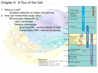
Microscopes vary in magnification and resolving power. Resolving power is a measure of image clarity. It is the minimum distance two points can be separated by and still be viewed as two separate points. Robert Hooke 1665
The minimum resolution of a light microscope is about 2 microns, the size of a small bacterium Light microscopes can magnify effectively to about 1,000 times the size of the actual specimen.
Techniques developed in the 20th century have enhanced contrast and enabled cell components to be labeled so that they stand out.
To resolve cell organelles we use an electron microscope (EM), which focuses a beam of electrons through the specimen or onto its surface. Electron microscopes have finer resolution than light microscopes
Transmission electron microscopes (TEMs) are used mainly to study the internal ultrastructure of cells. A TEM aims an electron beam through a thin section of the specimen. Cucumber cotyledon
Scanning electron microscopes (SEMs) are useful for studying surface structures. The image is focused on a screen Three dimensional The SEM has great depth of field, resulting in an image that seems three-dimensional. Rabbit trachea cells (SEM)
Electron microscopes reveal organelles, but they can only be used on dead cells. Light microscopes do not have as high a resolution, but they can be used to study live cells.
Cell fractionation separates the major organelles of the cells so that their individual functions can be studied. 2. Cell biologists can isolate organelles to study their functions and separate chemical components
This process is driven by an ultracentrifuge, a machine that can spin at up to 130,000 revolutions per minute and apply forces more than 1 million times gravity (1,000,000 g).
Microcentrifuge is standard equipment in biotechnology labs activities.
Equipment used to study cells at the genetic and protein level.
The Cell Theory Understanding the cellular nature of life followed the development of tools and techniques: In 1665, Robert Hooke observed "compartments" in a thin slice of cork (oak bark) using a light microscope. Used the term “Cell.”
By 1700, Anton van Leeuwenhoek developed simple light microscopes with high-quality lenses to observe tiny living organisms, such as those in pond water. "animalcules"
The Cell Theory Generalization that all living things are composed of cells. Cells are the basic unit of structure and function in living things Cells come from pre-existing cells
All cells are surrounded by a plasma membrane. All cells contain chromosomes which have genes in the form of DNA. All cells also have ribosomes 3. Two Major Classes of Cells: Prokaryotic and Eukaryotic Prokaryotic cell movie
Prokaryotic and eukaryotic cells differ in the location of chromosomes. • Eukaryotic cell chromosomes are in a nucleus. • In a prokaryotic cell, the DNA is concentrated in the nucleoid without a membrane separating it from the rest of the cell. Eukaryotic cell movie
The prokaryotic cell is much simpler in structure, lacking a nucleus and the other membrane-enclosed organelles of the eukaryotic cell.
What limits cell size? As a cell increases in size its volume increases faster than its surface area. Smaller objects have a greater ratio of surface area to volume. Square/Cube Law What cell organelle is critical in maintaining this ratio?
The plasma membrane functions as a selective barrier that allows passage of oxygen, nutrients, and wastes for the whole volume of the cell.
The volume of cytoplasm determines the need for this exchange. Rates of chemical exchange may be inadequate to maintain a cell with a very large cytoplasm. The need for a surface sufficiently large to accommodate the volume explains the microscopic size of most cells. Larger organisms do not generally have larger cells than smaller organisms - simply more cells.
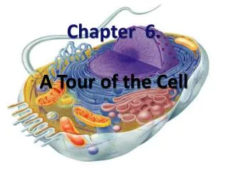
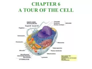
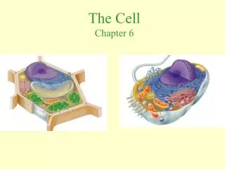
A eukaryotic cell has extensive and elaborate internal membranes, which partition the cell into compartments. Many enzymes are built into membranes. Membranes provide different local environments for specific metabolic functions. Each type of membrane has a unique combination of lipids and proteins for its specific functions. 4. Internal membranes compartmentalize the functions of a eukaryotic cell
The nucleus contains most of the genes in a eukaryotic cell. Some genes are located in mitochondria and chloroplasts. The nucleus is separated from the cytoplasm by a double membrane. Pores allows large macromolecules and particles to pass through. 5. The nucleus contains a eukaryotic cell’s genetic library
The nuclear side of the envelope is lined by a network of filaments that maintain the shape of the nucleus.
Within the nucleus, the DNA and associated proteins are organized into chromatin. In a normal cell they appear as a diffuse mass. When the cell prepares to divide, the chromatin fibers coil up to be seen as separate structures, chromosomes. What is special about chromosome numbers?
In the nucleus is the nucleolus. In the nucleolus, ribosomal RNA is synthesized and assembled with proteins to form ribosomal subunits. The subunits pass from the nuclear pores to the cytoplasm where they combine to form ribosomes.
Ribosomes contain rRNA and protein. A ribosome is composed of two subunits that combine to carry out protein synthesis. 6. Ribosomes build a cell’s proteins
What is implied if a cell type has large numbers of ribosomes and prominent nuclei. (e.g., pancreas) Free ribosomes, are suspended in the cytoplasm and synthesize proteins that function within the cytoplasm. Bound ribosomes, are attached to the outside of the endoplasmic reticulum.
The Endomembrane System • Many internal membranes in a eukaryotic cell are part of the endomembrane system. • The endomembrane system includes the nuclear envelope, endoplasmic reticulum, Golgi apparatus, lysosomes, vacuoles, and the plasma membrane. What is the adaptive value of this system?
The endoplasmic reticulum (ER) accounts for half the membranes in a eukaryotic cell. The ER includes membranous tubules and internal, fluid-filled spaces, the cisternae. 7. The endoplasmic reticulum manufactures membranes and modifies proteins
There are two regions of ER that differ in structure and function. Smooth ER looks smooth because it lacks ribosomes. Rough ER looks rough because ribosomes (bound ribosomes) are attached to the outside, including the outside of the nuclear envelope.
Smooth ER is rich in enzymes and plays a role in a variety of metabolic processes. Enzymes of smooth ER synthesize lipids, including oils, phospholipids, and steroids. The smooth ER helps catalyze conversion of glucose from stored glycogen in the liver. Smooth ER of the liver help detoxify drugs and poisons. (proliferation of smooth ER increases tolerance to the target and other drugs)
Rough ER is especially abundant in those cells that secrete proteins. As a polypeptide is synthesized by the ribosome, it is threaded into the cisternal space through a pore formed by a protein in the ER membrane. The protein is modified in the ER These secretory proteins are packaged in transport vesicles that carry them to their next stage.
Many transport vesicles from the ER travel to the Golgi apparatus for modification of their contents. The Golgi is a center of manufacturing, warehousing, sorting, and shipping. Which cells would have extensive Golgi apparatus? 8. The Golgi apparatus finishes, sorts, and ships cell products DR. CAMILLO GOLGI(1843-1926)
The Golgi apparatus consists of flattened membranous sacs - cisternae - looking like a stack of pita bread.
The lysosome is a membrane-bounded sac of hydrolytic enzymes that digests macromolecules. 9. Lysosomes are digestive compartments
Lysosomal enzymes can hydrolyze proteins, fats, polysaccharides, and nucleic acids. These enzymes work best at pH 5. What is the value of this compartmentalization?
The lysosomal enzymes and membrane are synthesized by rough ER and then transferred to the Golgi. At least some lysosomes bud from the trans face of the Golgi.
Lysosomes can fuse with food vacuoles, formed when a food item is brought into the cell by phagocytosis. Lysosomes can also fuse with another organelle or part of the cytosol. This recycling,or autophagy,renews the cell. Lysosome Movie
Vesicles and vacuoles (larger versions) are membrane-bound sacs with varied functions. Food vacuoles, from phagocytosis, fuse with lysosomes. Contractile vacuoles, found in freshwater protists, pump excess water out of the cell. Central vacuoles are found in many mature plant cells. 10. Vacuoles have diverse functions in cell maintenance