Download
1 / 106
1.06k likes | 1.31k Vues
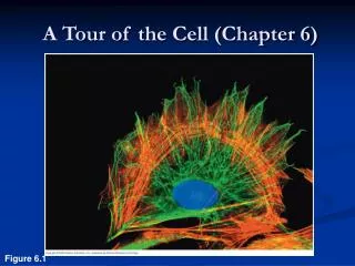
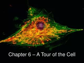
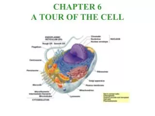
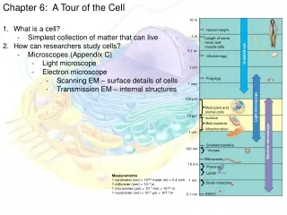
Chapter 6 A Tour of the Cell. Size of a Nanometer demo. http://learn.genetics.utah.edu/content/cells/scale/ Glucose = .9 nm Ribosome = 30 nm Mitochondria = 4000 nm long Skin Cell = 30,000 nm long = 30 um 1000 nm = 1 um. Light Microscope - LM.

E N D
Size of a Nanometer demo • http://learn.genetics.utah.edu/content/cells/scale/ • Glucose = .9 nm • Ribosome = 30 nm • Mitochondria = 4000 nm long • Skin Cell = 30,000 nm long = 30 um • 1000 nm = 1 um
Light Microscope - LM • Uses visible light to illuminate the object. • Relatively inexpensive type of microscope. • Can examine live or dead objects.
Resolution • Ability to detect two discrete points as separate from each other. • As Magnification increases, Resolution decreases. • LM working limits are100 - 1000X.
Limitations - LM • Miss many cell structures that are beyond the magnification of the light microscope. • Need other ways to make the observations.
Light Microscope Variations • Fluorescence: uses dyes to make parts of cells “glow”. • Phase-contrast: enhances contrasts in density. • Confocal: uses lasers and special optics to focus only narrow slides of cells.
After Before
Electron Microscopes • Use beams of electrons instead of light. • Invented in 1939, but not used much until after WWII. • Electron beam scans surface of the sample, exciting electrons which have their signals detected by a device that translates the pattern of electrons into an electronic signal to a video screen.
TEM – tracheal cell, long cilia were cut longitudinally, others cut to reveal cross section SEM rabbit trachea covered in cilia
Advantages • Much higher magnifications. • Magnifications of 50,000X or higher are possible. • Can get down to atomic level in some cases.
Disadvantages • Need a Vacuum. • High cost of equipment. • Specimen preparation. • Specimen must be dead
Other Tools for Cytology (study of Cells) • Cell Fractionation – break the cell apart and separate out the pieces based on their density.
History of Cells • Robert Hooke - Observed cells in cork. • Coined the term "cells” in 1665. • 1833 - Robert Brown, discovered the nucleus. • 1838 - M.J. Schleiden, all plants are made of cells. • 1839 - T. Schwann, all animals are made of cells.
Cell Theory • All living matter is composed of one or more cells. • The cell is the structural and functional unit of life. • All cells come from cells. • http://www.youtube.com/watch?v=mF9U5x6Nxnw
Types of Cells • Prokaryotic - lack a nucleus and other membrane bounded structures. • Eukaryotic - have a nucleus and other membrane bounded structures.
Both Have: • Membrane • Cytoplasm • Ribosomes (but the size is different)
Prokaryotic Eukaryotic Nucleus
Eukaryotic Prokaryotic
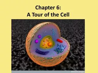
Basic Cell Organization • Membrane • Nucleus • Cytoplasm • Organelles
Membrane • Separates the cell from the environment. • Boundary layer for regulating the movement of materials in/out of a cell.
Cytoplasm or Cytosol • Cell substance between the cell membrane and the nucleus. • The “fluid” part of a cell. • Composed of water, dissolved salts and organic molecules (molecules that contain Carbon, dissolved proteins, and the cytoskeleton)
Organelle • Term means "small organ”. Formed body (or compartment) in a cell with a specialized function. • Important in organizational structure of cells.
Organelles - function • Way to form compartments in cells to separate chemical reactions. • Keeps various enzymes separated in space.
You must be able to: • Identify the major organelles • Give their structure • Give their function
Nucleus • Most conspicuous organelle. • Usually spherical, but can be lobed or irregular in shape.
Structure • Nuclear membrane • Nuclear pores • Nucleolus • Chromatin
Nuclear Membrane • Double membrane separated by a 20-40 nm space. • Inner membrane supported by a protein matrix which gives the shape to the nucleus.
Nuclear Pores • Regular “holes” through both membranes. • 100 nm in diameter. • Protein complex gives shape. • Allows materials in/out of nucleus.
Nucleolus • Dark staining area in the nucleus. • 0 - 4 per nucleus. • Ribosomes are made here
Chromatin • Chrom: colored • - tin: threads • DNA and Protein in a “loose” format. Will form the cell’s chromosomes.
Nucleus - Function • Control center for the cell. • Contains the genetic instructions.
Ribosomes • Structure: 2 subunits made of protein and rRNA. No membrane. • Function: protein synthesis.
Subunits • Large: • 45 proteins • 3 rRNA molecules • Small: • 23 proteins • 1 rRNA molecule
Locations • Free in the cytoplasm - make proteins for use in cytosol. • Membrane bound - make proteins that are exported from the cell.
Endomembrane System • Membranes that are related through direct physical continuity or by the transfer of membrane segments called vesicles.
Endoplasmic Reticulum • Often referred to as ER. • Makes up to 1/2 of the total membrane in cells. • Often continuous with the nuclear membrane.
Structure of ER • Folded sheets or tubes of membranes. • Very “fluid” in structure with the membranes constantly changing size and shape.
Types of ER • Smooth ER: no ribosomes. • Used for lipid synthesis, carbohydrate storage, detoxification of poisons/drugs. Stores Ca ions • Rough ER: with ribosomes. • Makes secretory proteins.
Golgi Apparatus • Structure: parallel array of flattened cisternae. (looks like a stack of Pita bread) • 3 to 20 per cell. • Likely an outgrowth of the ER system.