Download
1 / 42
430 likes | 676 Vues
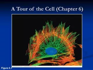
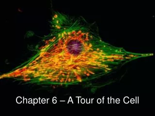
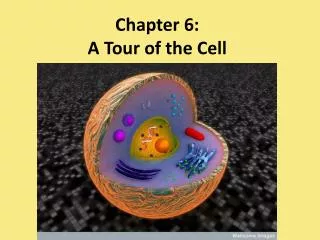
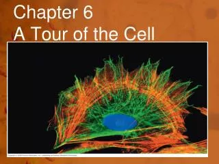
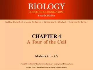
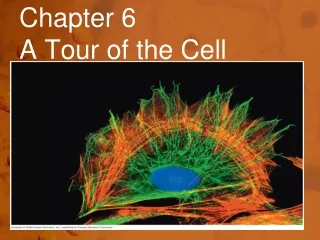
Chapter 6. A Tour of the Cell. Why do we study cells?. Cell Theory. All organisms are made up of cells The cell is the basic living unit of organization for all organisms All cells come from pre-existing cells. Biological diversity & unity.

E N D
Cell Theory • All organisms are made up of cells • The cell is the basic living unit of organization for all organisms • All cells come from pre-existing cells
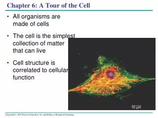
Biological diversity & unity • Underlying the diversity of life is a striking unity • DNA is universal genetic language • Cells are the basic units of structure & function • lowest level of structure capable of performing all activities of life
Activities of life • Most everything you think of a whole organism needing to do, must be done at the cellular level… • reproduction • growth & development • energy utilization • response to the environment • homeostasis
Cell characteristics All cells: • surrounded by a plasma membrane • have cytosol • semi-fluid substance within the membrane • cytoplasm = cytosol + organelles • contain chromosomes which have genes in the form of DNA • have ribosomes • tiny “organelles” that make proteins using instructions contained in genes Chromosomes
Types of cells • Prokaryotic vs. eukaryotic cells • Location of chromosomes Prokaryotic cell • DNA in nucleoidregion, without a membrane separating it from rest of cell Eukaryotic cell • chromosomes in nucleus, membrane-enclosed organelle
The prokaryotic cell is much simpler in structure, lacking a nucleus and the other membrane-enclosed organelles of the eukaryotic cell.
Eukaryotic cells • Eukaryotic cells are more complex than prokaryotic cells • within cytoplasm is a variety of membrane-bounded organelles • specialized structures in form & function • Eukaryotic cells are generally bigger than prokaryotic cells
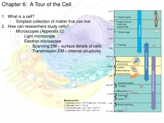
Limits to cell size Lower limit • smallest bacteria, mycoplasmas • 0.1 to 1.0 micron (µm = micrometer) • most bacteria • 1-10 microns Upper limit • eukaryotic cells • 10-100 microns • Giraffe nerve cells, ostrich egg are the largest cells • micron = micrometer = 1/1,000,000 meter • diameter of human hair = ~20 microns
What limits cell size? • Surface to volume ratio • as cell gets bigger its volume increases faster than its surface area • smaller objects have greaterratio of surface area to volume Surface area = 96 mm2 Volume = 64 mm3 Why is a huge single-cell creature not possible?
Limits to cell size • Metabolic requirements set upper limit • in large cell, cannot move material in & out of cell fast enough to support life aa aa What process is this? CH NH3 aa CHO O2 CH CHO CO2 CHO CO2 CO2 aa NH3 O2 NH3 O2 NH3 CHO aa CO2 CH aa CH O2 aa O2
How to get bigger? But what challenges do you have to solve now? • Become multi-cellular (cell divides) CO2 CO2 O2 NH3 aa NH3 aa CO2 NH3 O2 CO2 CO2 CH CHO CO2 NH3 aa O2 NH3 NH3 CO2 CO2 CO2 CHO aa NH3 NH3 NH3 CH CHO CO2 CO2 O2 aa aa CH
Cell membrane • Exchange organelle • plasma membranefunctions as a selective barrier • allows passage of O2, nutrients & wastes
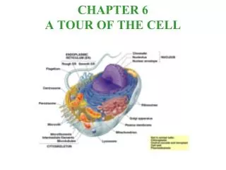
Organelles & Internal membranes • Eukaryotic cell • internal membranes • partition cell into compartments • create different local environments • compartmentalize functions • membranes for different compartments are specialized for their function • different structures for specific functions • unique combination of lipids & proteins Mitochondrion Endoplasmic Reticulum Golgi apparatus Chloroplast
Nucleus • Chromatin & Chromosomes • Nucleolus • Nuclear Envelope • Ribosomes • Endoplasmic Reticulum (smooth & rough) • Golgi Apparatus • Lysosome • Food vacuoles • Contractile vacuoles • Central Vacuole • Mitochondria • Chloroplasts • Peroxisome • Cytoskeleton • Centrosomes • Centrioles • Cilia & Flagella • Cell wall • Microfilaments • Microtubules • Plasmodesmata Cell Organelles
Nucleus & Nuclear structure • Enclosed by nuclear envelope (double membraned) w/ nucleopores • Internally lined with nuclear lamina (network of protein) for support & shape • Nuclear matrix fibers internally • Chromatin (uncoiled DNA & protein) then become chromosomes • Nucleolus (no membrane) produces rRNA & then ribosomes
Ribosomes Found in Eukaryotic and Prokaryotic cells Made of rRNA and protein Carry out Protein Synthesis Human pancreas cell has a few million Not membrane bound Free or bound to Rough Endoplasmic Reticulum Free produce enzymes for glycolysis of glucose Bound to Endoplasmic Reticulum for protein synthesis
Endomembrane system • Series of membranes for specific tasks • Endoplasmic reticulum (ER) – network of membranes & sacs called cisternae. Continuous w/ nuclear envelope • Smooth – synthesis of lipids, metabolism of carbohydrates, detoxification of drugs & poisons & storage of Calcium ions • Rough – studded w/ ribosomes for protein synthesis. • Transport vesicles – sacs containing proteins that break free from RER to be transported to another cell
Golgi Apparatus Golgi Apparatus song Shipping & receiving center Products of ER are modified, packaged & shipped to other parts of the cell or to other cells Flattened cisternae Vesicles on ends for transport bud off Cis side for receiving and trans for shipping
Lysosomes • Membrane bound sacs containing hydrolytic enzymes • Animal cells only • Acidic for optimal enzyme activity • Autodigestion (autolysis) when lysosomes break open (cleans up dead cells) or autophagy (cleans up non-functioning organelles) • Fuse with food vacuoles for digestion • Tay-Sach’s disease – inheritable – lysosomes do not make enzymes
Vacuoles • Food vacuoles formed by phagocytosis • Contractile vacuoles – like sump pumps to control osmotic pressure in freshwater protists • Central vacuole in plants enclosed by tonoplast– holds reserve organic compounds, K and Chloride, stores metabolic byproducts, may contain defensive toxins, stores water reserve to maintain structure (turgor pressure)
MITOCHONDRIA & Chloroplasts • Both involved in energy conversion • Mitochondria– site of cellular respiration • 1 – 10 um long (same size as bacteria!) Also can move • Double membrane • Inner membrane folds in – Cristae(> surface area) • Electron Transport Chain of respiration occurs here • Mitochondrial matrix – inner portion – Krebs Cycle • Highest in human muscles and liver cells • Contains own DNA so can be replicated as needed
Plastids • Amyloplasts– store starch • Chromoplasts – store flower & fruit pigments • Chloroplasts – contain chlorophyll & enzymes involved in photosynthesis • 2 – 5 um with DNA (same as bacteria!!) • At least double membraned • Granum – interconnected membrane stacks of individual thylakoidswith fluid called stroma
Peroxisomes • Single membrane • Contain enzymes to transfer H to O2 to produce H2O2for fatty acid breakdown for fuel or for the liver to detoxify alcohol • Also contains catalase & superoxide dismutase to break down hydrogen peroxide
Cytoskeleton • Network of fibers throughout the cytoplasm • Provides support (especially in animal cells) • Cell Motility – motor proteins • Cilia & flagella motion • Microfilaments and muscle contractions • Moves vesicles around cells • Streaming of cytoplasm - cyclosis
Components of Cytoskeleton • Microtubules - thickest • Microfilaments – thinnest • Intermediate filaments – middle range diameter
Microtubules • Hollow rods (~25nm in diameter) & 200nm – 25um in length • Wall is made of protein tubulin • Shape & support the cell • Tracks for motor proteins to travel along • Form centrioles (9+3 organization)in animal cells for cell division
Cilia and Flagella • Specialized collection of microtubules for movement of cilia & flagella • Cilia & Flagella • Cilia are found in large #’s & are small - .25um in diameter to 2 – 20un in length • Move like oars w/ power & recovery strokes • Flagella usually single to few and longer – 10 – 200um • Undulating motion or as a propeller • Animation
Structure of Cilia & Flagella • 9 + 2 microtubule organization • Proteins cross link pairs to central pair • Anchored by basal body • Dynein– protein connecting one pair onto next pair. • Responsible for bending movements of cilia/flagella
Microfilaments: Actin fibers • Solid ~ 7nm rods across • Actin – globular proteins • Twisted double chain of actin subunits • Form structural networks throughout cytoplasm • 3D network inside cell membrane • Make up microvilli in intestines • Along with myosin, makes up muscles
Muscle movement • Myosin acts as motor protein along actin filaments • Myosin “walks” along actin • Also involved in amoeboid motion and formation of pseudopods and cytoplasmic streaming in plant cells
Intermediate Filaments • 8 – 12nm diameter • More permanent structures in cytoskeleton • Reinforce shape of cell and “fix” organelle positions
Extracellular Components & Connections • Cell Wall in plant, fungi, bacteria and some protist cells aka, every cell but animals & some protist • Protection • Structure • Prevents excess uptake of water • Supports plant against gravity • Range in size from 0.1um to several nm • Composition varies from species to species
Structure of plant Cell Wall • Primary cell wall – first to be produced, flexible & thin • Middle Lamella of pectin – thin sticky polysaccharide to hold layers together (glue) • Secondary cell wall forms when plant is mature & stops growing. More rigid • Plasmodesmata perforate cell walls and form channels
Animal Cell Extracellular Matrix (ECM) • ECM made up of glycoproteins including collagen – strong fibers outside cell • Collagen accounts for ½ total body proteins • Collagen fibers embedded in Proteoglycan molecules • Fibronectin (purple rods) attached EMC tointegrinsthat bind to plasma membrane • Involved in communications between cells Fibronectin
Intracellular connections • Plasmodesmata - Connects plant cells to other plant cells. • Tight junctions (form seals like a tongue and groove joint) • Desmosomes(act like rivets) • Gap junctions (act like channels) found in epithelial cells