Download
1 / 11
110 likes | 120 Vues
Learn about the integumentary system and body membranes, including cutaneous, serous, and mucous membranes. Explore the skin's functions, components, and structure, from the epidermis to the dermis and subcutaneous layer. Delve into skin appendages like hair, nails, and glands, understanding their roles, production, and functions.

E N D
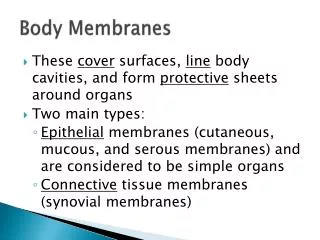
Epithelial Membranes: epithelium and its underlying connective tissue • Cutaneous Membranes – skin • Serous Membranes – simple squamous on connective tissue basement membranes, lines body cavities. • Parietal – lines walls of cavities • Visceral – covers organs (silver skin on red meat) 3. Mucous Membranes – line surfaces open to exterior, produce thick mucous to soften and moisten.
The Skin Functions: • Protection – infection barrier • Temperature Regulation – cools with sweat glands • Sense Organ Activity – via nerve endings and specialized receptors
The skin cont.Components in 1 square inch of Skin • 500 sweat glands • 1000 nerve endings • Yards of blood vessels • 100 oil glands • 150 pressure sensors • 75 heat sensors • 10 cold sensors • Millions of cells
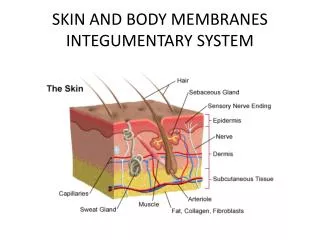
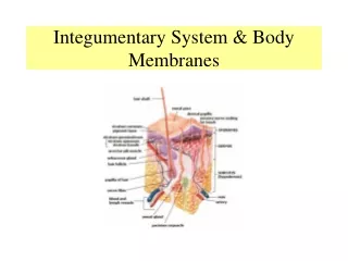
The skin cont.Structure of skin (See pg. 123) Subcutaneous Layer or Hypodermis • Loose connective tissue and fat • Insulates • Stores energy • Food for muscle • Shock absorbing
The skin cont.Structure of skin (See pg. 123) Epidermis – outer most layer • Stratum Germinativum – site of mitosis, cells cytoplasm replaced by keratin • Stratum Corneum – dead cells • Pigment Layer – melanocytes with melanin, layers spot welded together which leads to blisters.
The skin cont.Structure of skin (See pg. 123) Dermis • Deeper and thicker • Scattered cells with fibers in-between • Dermal Papillae – parallel peg-like rows that bind layers together and make up finger print • Location of Accessory organs such as: • Muscle fiber • Hair follicle • Sweat and Sebaceous Glands • Blood vessels
Appendages of Skin (still on pg. 123) Hair • Hair follicle – epidermal and tube-like, grows into dermis • Hair Papilla – beginning point of growth • Root – hidden in follicle • Shaft – visible part of hair • Arrector pili – muscle connected to follicle, causes goose bumps
Appendages of Skin (still on pg. 123) Continued Receptors • Meissners Corpuscle – near surface, light touch • Pacinian Corpuscle – deep in dermis, pressure • Krauses End Bulbs – sense cold • Organ of Ruffini – sense heat • Nerve Endings – sense pain
Appendages of Skin (still on pg. 123) Continued Nails • Produced in the epidermis • Nail Body – visible • Root – hidden • Cuticle – hides root • Lanula – near root, “half-moon”
Appendages of Skin (still on pg. 123) Continued Glands • Sweat or Sudoriferous Glands • 3 mill plus, produce 12l every 24 hours • Eccrine – numerous and important in producing sweat, eliminating waste and regulating body temp. • Apocrine – in armpits and genitals, produce thicker milky secretions, susceptible to bacterial contamination. • Sebaceous– produces oil for hair and skin, increases during adolescence, clogged ducts = white heads, when aged and darkened = black head. • Acne – sebaceous ducts filled with cells and sebum