Download
1 / 97
1.01k likes | 1.35k Vues
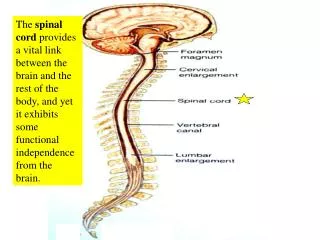
The spinal cord provides a vital link between the brain and the rest of the body, and yet it exhibits some functional independence from the brain. The adult spinal cord travels from the foramen magnum and terminates within the vertebral foramen of the first lumbar vertebra (L1) in adults.

E N D
The spinal cord provides a vital link between the brain and the rest of the body, and yet it exhibits some functional independence from the brain.
The adult spinal cord travels from the foramen magnum and terminates within the vertebral foramen of the first lumbar vertebra (L1) in adults.
The spinal cord can be subdivided into five regions: cervical region, thoracic region, lumbar region, sacral region, and coccygeal region (which has only one pair of nerves). Don’t be confused and think that the sacral “region” of the spinal cord is surrounded by sacral vertebrae. It is NOT!
The diameter of the spinal cord is the largest in the cervical region and there is a larger proportion of white matter compared to gray matter.
The diameter of the sacral region of the spinal cord (which is surrounded by the T12/L1 vertebrae) is the smallest and the proportion of gray matter is largest in the spinal cord.
The cervical enlargement contains the neurons that innervate the upper limbs The lumbar enlargement contains the neurons that innervate the lower limbs.
The tapering end of the spinal cord is called the conus medullaris. The conus medullaris is surrounded by L1 in and adult and L2 in a child.
The adult spinal cord terminates at the level of the first lumbar vertebra (L1) In a developing child, the spinal cord can extend to the level of the second lumbar vertebra (L2)
The cauda equina (horse’s tail) is composed of nerves that arise from the conus medullaris and extend inferiorly.
The filum terminale, which is composed of pia mater, extends from the conus medullaris to the coccyx. Note the subarachnoid space also continues for some distance.
There are 31 pairs of spinal nerves that serve defined segments of the human body.
There are 8 pairs of cervical spinal nerves. This is possible because the first pair (C1 spinal nerves) exits the spinal column between the occipital bone and the atlas (C1). The remaining 7 pairs (C2-C8 spinal nerves) exit below each of the 7 cervical vertebrae via the intervertebral foramina. All the spinal nerves are mixed nerves.
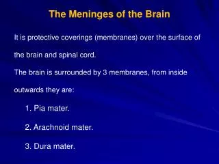
The spinal cord is surrounded by the dura, arachnoid, and pia maters (the meninges)
WHAT IS THE NAME OF THE NERVE THAT EXITS VIA THE INTERVERTEBRAL FORAMEN BETWEEN THE ATLAS AND THE AXIS? A VAGUS NERVE B FIRST CERVICAL SPINAL NERVE C ACCESSORY NERVE D LONG THORACIC NERVE E SPINAL NERVE C2
The epidural space is between the vertebra and the dura mater
The dura mater extends along the entire length of the vertebral canal and surrounds the spinal cord. It also extends along the initial portion of the radiating spinal nerves
In this midsagittal picture #3 is the dura mater, #5 is the spinal cord, # 4 is the epidural space, and #6 is the subarachnoid space where CSF is located (#1 is an intervertebral disc and #2 is the body of a vertebrae).
Spinal taps are done between the third and fourth lumbar vertebrae because there is no spinal cord at that location
The tip of the needle is inserted into the subarachnoid space outside the cauda equina and spinal fluid is removed for testing.
The entering pressure can be determined when the needle is inserted into the subarachnoid space during a spinal tap.
Spinal fluid is normally crystal clear like water. Cloudy spinal fluid, like the specimen shown, is a sign of white blood cells (pus). The most common cause for white blood cells in the spinal fluid is viral or bacterial meningitis.
WHICH OF THE FOLLOWING IS TYPICALLY PENETRATED DURING A ROUTINE SPINAL TAP? A PIA MATER B NUCLEUS PULPOSUS C ANULUS FIBROSUS D SPINAL CORD E NONE OF THE ABOVE
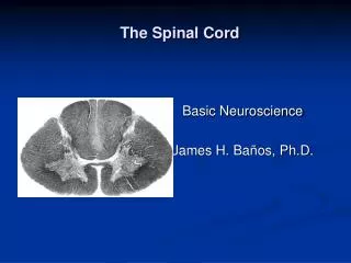
The cross-sectional view shows that the gray matter is central and the white matter is peripheral
The peripheral white matter contains ascending and descending tracts of nerves traveling to and from the brain. The central gray matter serves as a center for spinal reflexes.
The central canal runs the entire length of the spinal cord, is contiguous with the brain and contains cerebrospinal fluid (CSF)
The spinal cord develops as 31 segments, each of which gives rise to a pair of spinal nerves that emerge from the cord through the intervertebral foraminae
Mixed nerves carry both types of information and some axons are transmitting impulses in one direction, while other axons are transmitting impulses in the opposite direction. All spinal nerves are mixed nerves.
There are 8 pairs of cervical spinal nerves. This is possible because the first pair (C1 spinal nerves) exits the spinal column between the occipital bone and the atlas (C1). The remaining 7 pairs (C2-C8 spinal nerves) exit below each of the 7 cervical vertebrae via the intervertebral foramina. All the spinal nerves are mixed nerves.
Most of the spinal nerves are associated with specific dermatomes (an area of skin innervated by all the cutaneous neurons of a certain spinal or cranial nerve).
trigeminal Dermatome map. Note the trigeminal nerve has dermatomes on the face.
Dermatomes of the trigeminal nerve (cranial nerve V) are seen on the face
Note that the trigeminal nerve has dermatomes on the face (see white area) and that the first pair of cervical spinal nerves (C1 spinal nerves) are not represented on the surface at all.
Chickenpox (varicella) virus is acquired by the respiratory route and causes a head-to-toe rash in children. The chickenpox virus can invade the ganglia along the spinal cord and remain latent until adulthood. It can then be activated by suppression of the immune system. It will then travel through sensory axons of a single dermatome and erupt onto the skin in a single dermatome on one side of the body (unilateral eruption)
Shingles is a reactivation of latent chickenpox from childhood that travels to the surface via a single nerve on one side of the body.
Shingles involving the first (ophthalmic) division of the trigeminal nerve (cranial nerve V) on face.
Explanation of referred pain. Numerous cutaneous and visceral sensory neurons share the same ascending tracts.
WHICH OF THE FOLLOWING IS CORRECT ABOUT SHINGLES? A ADULTS WITH THIS CONDITION CAN CAUSE A HEAD-TO-TOE RASH IN CHILDREN B IT TYPICALLY OCCURS BILATERALLY C IT IS TRIGGERED BY ETHYL ALCOHOL AND PROLONGED NERVE COMPRESSION D IT IS MOST COMMON IN PERSONS UNDER 50 E ALL OF THE ABOVE
The majority of the spinal nerves combine and then split again as networks of nerves referred to as plexuses. The exceptions are T2-T12 and S5-Co1, which do NOT form plexuses
The cervical plexus is formed primarily by spinal nerves C1-C4 (C5 is not considered part of this plexus, even though it contributes some axons)
The cervical plexus, and particularly spinal nerves C3, C4, and part of C5, give rise to the phrenic nerve which innervates the diaphragm. Injury above C3 would lead to death by suffocation.