Download

1 / 41
450 likes | 901 Vues
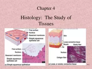
HISTOLOGY & EMBRYOLOGY Teaching PPT Dept. of Anat., Hist. & Embry. School of Medicine Xi’an Jiaotong University. HUMAN EMBRYOLOGY. Development of Urinary & Genital systems. Tianbao Song ( 宋 天 保 ). Chapter 26 Development of Urinary and Genital System I . Development of Urinary System.

E N D
HISTOLOGY & EMBRYOLOGY Teaching PPT Dept. of Anat., Hist. & Embry. School of Medicine Xi’an Jiaotong University
HUMANEMBRYOLOGY Development of Urinary & Genital systems Tianbao Song (宋 天 保)
Chapter 26 Development of Urinary and Genital System I. Development of Urinary System 1. Development of Kidney and Ureter • 1.1 Primordia: Intermediate mesoderm: • Cervical part → nephrotomes • Caudal part →nephrogenic cords.
1.2 Pronephros: Nephrotomes→ pronephric tubules and duct→ pronephros → degenerates. 1.3 Mesonephros: 1) Nephrogenic cord →mesonephric ridge→ mesonephros
2) Mesonephric tubules → mostdisappear; • mesonephric duct→ open into cloaca.
1.4 metanephros 1) Primordia: ureteric bud and metanephric blastema
Mesonephric duct 2)ureteric bud→ ureters,renal pelvis, calyces, collecting tubules
3) Metanephric blastema→nephrons →Bowman's capsule, renal tubules → open connection to collecting tubules
2. Formation of Bladder and Urethra Primordium: urogenital sinus 1) upper part→urinary bladder→ continuous with allantois (obliterated later)
2)Middle part of urogenital sinus →urethra; 3)Lower part→ penile urethra in males, vestibule in females.
3. Congenital Malformations • 3.1 Polycystic kidney • Abnormal development of the collecting system, or failure of the collecting tubules and nephrons to join; • Kidney contains many cysts, and failure of renal function may be caused.
3.2 Pelvic kidney • Failure of kidney to ascend and still in the pelvis. • 3.3 Horseshoe kidney • Both kidneys fail to ascend, and their lower poles fuse together.
3.4Double ureter • Early splitting of ureter completely or partially; • Ureters open into bladder separately, or unite and open as usual.
3.5 Urachal fistula • Caused by persisting allantois. • Urine may drain from the umbilicus.
II. Development of Genital System 1. Development of Gonads 1.1 Primordia: gonadal ridges(coelomic epithelium + mesenchyme),primordial germ cells.
1.2 Indifferent stage: • Gonadal ridges→ • primary sex cords. • Yolk sac→ primordial • germ cells migrate into • primary sex cords
1.3 Development of testis 1) Y→ SRY→ primary sex cords → medulla → testicular cords →seminiferous tubules→ ↗epithelial cells → Sertoli cells ↘primordial germ cells → spermatogonia
2) Mesenchyme (surface) →tunica albuginea; (between sex cords ) →Leydig cells→ (androgens)
1.4 Development of ovary 1) XX(no SRY) → primary sex cords → medulla → degenerate 2) Coelomic epithelium→ secondary sex cords (with primordial germ cells )
3) Secondary sex cords → cell clusters → • germ cells → oogonia • epithelial cells → follicular cells Primordial follicles 4) Oogonia → primary oocyte →no oogoniaat birth.
1.5 Descent of the testis 1) Mesenchyme → gubernaculums (testis -- genital swelling)
2) Rapid body growth gubernaculum shortening → testis descent→ inguinal canal→ scrotum
3) Peritoneal sac →vaginal process→ scrotum → tunica vaginalis 4) Proximal part of vaginal process → obliterated at birth.
2. Development of Genital Duct 2.1 Primordia: mesonephric duct,paramesonephricduct.
2.2 Indifferent stage • Coelomic epithelium→ • paramesonephric ducts: • cranial end → body cavity; • caudal ends fuse → • uterinecanal. • 2) Tip of uterine canal → • urogenital sinus → sinus • tubercle.
2.3 Development of male genital duct 1) Androgens (Leydig cells)→ • mesonephric duct→ ductus epididymis, ductus deferens, ejaculatoryduct, seminal gland; • mesonephric tubules→ efferent ductules of testis.
2) Anti-Mullerian duct hormone (Sertoli cells)→paramesonephric ducts regress.
2.4 Development of female genital duct (no A & AMH) 1)Paramesonephric duct→ uterine tube; Uterine canal→uterus, upper 1/3 of vagina.
2)Sinus tubercle→vaginal plate→ canalized → lower 2/3 of vagina. 3) Mesonephric ducts → degenerate.
3. Development of external genitalia 3.1 Primordia: urogenital folds, genital tubercle, urogenital groove, labioscrotal swellings.
3.2 Development • Male (A) Female(no A) • genital tubercle phallus clitoris • urogenital folds lateral wall of urethra labia minora • labioscrotal swellings scrotum labia majora • urogenital groove penile urethra vestibule
4. Congenital Malformations • 4.1 Cryptorchidism • Failure of one or both testes to descendinto scrotum; • Seeming to be due to abnormal androgen production; • Testes may remain in abdomen or in inguinal canal.
4.2 Congenital inguinal hernia • Failure of vaginal process to close; • Intestinal loops may descend into scrotum.
4.3 Abnormalities of the uterus • Defects of fusion of caudal ends of paramesonephric ducts; • May cause double uterus, bicornuate uterus, uterus septus, etc.
4.4 Vaginal atresia Caused by failure of vaginal plate to form or to be canalized.
4.5 Hermaphroditism 1)True hermaphrodite has gonad and external genitalia of both sexes. rarely observed. • 2)Pseudohermaphrodite • Has either testes (male) or ovaries (female); • External genitalia resembling opposite sex; • Inadequate (male) or excessive (female) androgen production.
4.6 Testicular feminization syndrome • 44+XY chromosome complement; • Devoid of androgen receptors; • Testes in inguinal region, no spermatogenesis; • External genitaliaas in females; no uterine tubes, uterus.
4.7Hypospadias • Incomplete fusion of urogenital folds; • Abnormal opening of urethra along ventral penis.
SUMMARY • Primordia and developmental featuresof gonads & kidneys. • Sex differentiation of the gonads. 2. • Differentiation of the urogenital sinus; • 4. Sex differentiation of the genital ducts. • 5. Congenital malformations of genital system.