Analytical X-ray Diffraction Safety Training Part I
680 likes | 831 Vues
Analytical X-ray Diffraction Safety Training Part I. Slides developed by John Pickering SJSU Radiation Safety Officer (RSO) (Retired). What is the purpose of radiation safety training?.

Analytical X-ray Diffraction Safety Training Part I
E N D
Presentation Transcript
Analytical X-ray Diffraction Safety TrainingPart I Slides developed by John PickeringSJSU Radiation Safety Officer (RSO) (Retired)
What is the purpose of radiation safety training? To help you gain enough knowledge to enable you to perform your job safely. To ensure that you adhere to proper radiation protection practices while working with or around x-ray generating devices.
Fundamental Radiation Physics • Radiation – alpha particles, beta particles, gamma rays, X-rays • Radioactivity – spontaneous nuclear transformations • Generally alpha particles and beta particles • Often accompanied by gamma ray emission • Ionizing Radiation – radiation capable of producing charged particles (ions) in the material through which it passes
Radiation is energy in transit in the form of high speed particles and electromagnetic waves. We encounter electromagnetic waves every day. They make up our visible light, radio and television waves, ultra violet (UV), and microwaves with a spectrum of energies. These examples of electromagnetic waves do not cause ionization of atoms because they do not carry enough energy to remove electrons from atoms.
Ionizing radiation Ionizing radiation is radiation with enough energy so that during an interaction with an atom, it can remove tightly bound electrons from their orbits, causing the atom to become charged or ionized. Ionizing radiation deposits energy at the molecular level, causing chemical changes which lead to biological changes. These include cell death, cell transformation, and damage which cells cannot repair. Effects are not due to heating.
Radiation Units • Roentgen (R)The roentgen (R) is a unit of radiation exposure in air. • It is defined as the amount of x-ray or g radiation that will generate 2.58E-4 coulombs/kg of air at standard temp and pressure. • radRAD stands for Radiation Absorbed Dose and is the amount of radiation that will deposit 0.01 J/kg of material. • A roentgen in air can be approximated by 0.87 rad in air, 0.93 rad in tissue, and 0.97 rad in bone. • Dose • The SI unit of absorbed dose is the gray (Gy), which has the units of J/kg. 1 Gy= 100 rad.
Radiation Units • REMREM stands for Roentgen Equivalent Man. The REM is a unit of absorbed dose and is equal to the rad multiplied by a weighting factor which varies according to the type of radiation. The weighting factor for x-rays is equal to 1. • For x-rays, one rem is equal to one rad. • The SI unit used in place of the rem is the sievert (Sv). 1 Sv = 100 rem.
Radiological Fundamentals The basic unit of matter is the atom. Nucleus Electron Nucleus Neutrons Protons
X-RAY AND GAMMA ( ) RAY PROPERTIES Charge: None Mass: None Velocity: 3 x 10⁸ m/s Origin: Rays: Nucleus X Rays: Electron Cloud & Bremsstrahlung
What are x-rays? • X-rays are photons (electromagnetic radiation) which originate in the energy shells of an atom, as opposed to gamma rays, which are produced in the nucleus of an atom.
Background Radiation Cosmic - 28 mrem Radon - 200 mrem Diet - 40 mrem Terrestrial - 28 mrem
Man-made sources of radiation contribute to the annual radiation dose (mrem/yr). Round trip US by air 5 mrem per trip Building materials - 3.6 Gas range - 0.2 Smoke detectors - 0.0001 Man-made Radiation Cigarette smoking - 1300 Medical - 53 Fallout < 1
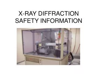
Radiation Sources • X-ray diffraction is a source of very intense radiation. • The primary beam can deliver as much as 400,000 R/minute • Collimated and filtered beams can produce about 5,000 to 50,000 R/minute • Diffracted beams can be as high as 80 R/hour
Dose Limits • EPA Guidance for dose limits • NRC Regulations for dose limits • DOE Regulations for dose limits • DOT Regulations for transport • State Agreement States • NCRP National Scientific Body • Licensee Institutional Admin Limits
Radiation Worker Whole Body Extremities Skin and other organs Lens of the eye Non-Radiation Worker Embryo/fetus Visitors and Public 5 rem/year - 3 rem/quarter 50 rem/year 50 rem/year 15 rem/year 0.5 rem/year 0.5 rem/gestation period 0.1 rem/year Regulatory Limits
ALARA Program • As Low As Reasonably Achievable • Responsibility of all employees • Exposures shall be maintained ALARA • Below regulatory limits • No exposure without commensurate benefit
Responsibilities for ALARA Ultimately YOU are! • To establish a program • Meet regulatory limits Management Safety Organization • Implementing a program • Run the daily operation Radiation Worker • To follow program
General Methods of Protection • Time • Distance • Shielding
What are x-rays? • X-rays are produced when accelerated electrons interact with a target, usually a metal absorber, or with a crystalline structure. This method of x-ray production is known as bremsstrahlung. • The bremsstrahlung produced is proportional to the square of the energy of the accelerated electrons used to produce it, and is also proportional to the atomic number (Z) of the target (absorber).
What are x-rays? • Many different types of machines produce x-rays, either intentionally or inadvertently. Some devices that can produce x-rays are x-ray diffractometers, electron microscopes, and x-ray photoelectron spectrometers. • X-rays can also be produced by the attenuation of beta particles emitted from radionuclides.
How X-rays are Produced When fast-moving electrons slam into a metal object, x-rays are produced. The kinetic energy of the electron is transformed into electromagnetic energy. X-ray Tube
What are X-rays • Electromagnetic radiation • Originate in energy shells of atom • Produced when electrons interact with a target target electron X-ray
Characteristic X-rays Characteristic x-rays are produced by transitions of orbital electrons from outer to inner shells. Since the electron binding energy for every element is different, the characteristic x-rays produced in the various elements are also different. This type of x-radiation is called characteristic radiation because it is characteristic of the target element. The effective energy characteristic x-rays increases with increasing atomic number of the target element.
Bremsstrahlung Radiation • A projectile electron that completely avoids the orbital electrons on passing through an atom of the target may come sufficiently close to the nucleus of the atom to come under its influence. • Since the electron is negatively charged and the nucleus is positively charged, there is an electrostatic force of attraction between them. • As the projectile electron approaches the nucleus, it is influenced by a nuclear force much stronger than the electrostatic attraction. • As it passes by the nucleus, it is slowed down and deviated in its course, leaving with reduced kinetic energy in a different direction. • This loss in kinetic energy reappears as an x-ray photon.
Bremsstrahlung Radiation Z2 A Bremsstrahlung production =
Photon Energy and Total Power As the voltage increases the penetration increases As the Current increases the dose rate increases Dose (Current) Energy Voltage (Penetration) The total power W = V x A
Photon Energy and Total Power Characteristic X-ray Gamma Peak (Specific Energy) Dose Energy
Photon Energy and Total Power Average Energy = 1/3 Maximum Energy Dose Energy
Photon Energy and Total Power Adding Filtration Filtration can shift the average Energy (voltage) higher Current Voltage (Penetration)
Interaction with Matter • When x-rays pass through any material • some will be transmitted • some will be absorbed • some will scatter • The proportions depend on the photon energy and type of material
Emission Radiation Emission
Absorption Absorption
Reflection Reflection
Skyshine Skyshine
X-ray Safety for Operators • Decrease dose to the operator • Time • Determines total dose • Voltage • Determines penetration • Current • Determines dose rate
Ionizing Radiation Produces damage through ionization and excitation
X-ray Safety Filtration removes low-energy x-rays from the primary beam. Collimation limits the beam to a useful area. Compliance testing performed periodically. Registration of sources with regulatory agency.
At HIGH Doses, We KNOW Radiation Causes Harm • High Dose effects seen in: • Radium dial painters • Early radiologists • Atomic bomb survivors • Populations near Chernobyl • Medical treatments • Criticality Accidents • In addition to radiation sickness, increased cancer rates were also evident from high level exposures.
Law of Bergonie and Tribondeau • The more rapidly reproducing cells are more radiosensitive. • The least functionally differentiated cells are more radiosensitive. 1903
Dividing Cells are the Most Radiosensitive • Rapidly dividing cells are more susceptible to radiation damage. • Examples of radiosensitive cells are • Blood forming cells • The intestinal lining • Hair follicles • A fetus This is why the fetus has an exposure limit (over gestation period) of 500 mrem (or 1/10th of the annual adult limit)
Biological Effects of Radiation • are dependent upon: • Total energy deposited • Distribution of deposited energy Low dose, low-dose rate radiation exposure. The effects are in great dispute. It is thought that the effects of a protracted dose of radiation are not as great as with an acute dose because of biological repair mechanisms.
Relative Radiosensitivity ofMammalian Tissues Sensitive • Spermatogonia • Lymphocytes Hematopoietic Tissues Less sensitive • Epithelium • Epidermus Resistant • Central nervous system • Muscle • Bone