Aortic Stenosis
Aortic Stenosis. Introduction. Etiology/Classification Pathophysiology Clinical Manifestations Evaluation Management. Obstruction to left ventricular outflow. Valvular Calcific/Degenerative Biscuspid Rheumatic Congenital Supravalvular

Aortic Stenosis
E N D
Presentation Transcript
Introduction • Etiology/Classification • Pathophysiology • Clinical Manifestations • Evaluation • Management
Obstruction to left ventricular outflow • Valvular • Calcific/Degenerative • Biscuspid • Rheumatic • Congenital • Supravalvular • Uncommon. Occurs as part of a congenital syndrome or from lipid deposits in severe familial hyperlipidemias • Subvalvular • Congenital • Fibromuscular membrane present in the LVOT • Pathogenesis not completely understood • Hypertrophic Cardiomyopathy
Valvular Aortic Stenosis • Calcific/Degenerative • Most common • Normal trileaflet valve develops “age-related” calcification • Presents later in life (age 70-85) • Bicuspid valve • Present in 1-2% of the population • More prevalent in men (70-80% of cases) • Usually presents at a younger age (45-65) • Rheumatic • Preferentially involves the mitral valve, so Rheumatic AS diagnosed when occurs concurrently with mitral valve disease • Congenital • Typically unicuspid valve with single eccentric orifice • Ochronosis with alkaptonuria • Anyone?
Calcific/Degenerative AS • Active disease process • “Rather than representing a degenerative and unmodifiable process, valvular aortic stenosis is an active process initiated at least in part by mechanical injury, mediated by lipid infiltration and a chronic inflammatory reaction, and further characterized by accumulation of extracellular matrix secreted primarily by resident valve fibroblasts as a response to inflammatory injury.” (Circulation 1994;90:844-53) • Evidence that hemodynamic stress plays a role, as calcium deposition occurs on the aortic side of the valve where turbulence and shear stress are high.
Relation to atherosclerosis? • Some commonality in risk factors for CAD and AS • Statins may have a role in slowing the progression of AS • Increased temperature variability in valves of patients with AS (similar to previous studies looking at thermal examination of CAD plaques)
Normal • Normal anatomy: three thin cusps with unrestricted systolic opening • Normal area 3-4 cm² • Normal opening produces 2cm leaflet separation • Normal velocity: approx 1 m/s
Pathophysiology • Aortic sclerosis produces thickening of the valves, but no obstruction to outflow • When stenosis develops, the functional area of the valve decreases and causes a measurable obstruction of outflow • Concentric LVH develops with normal chamber size • Diastolic dysfunction due to increased myocardial cell mass and interstitial fibrosis • Atrial contraction plays an important role in filling of the left ventricle in AS • Loss of appropriately timed atrial contraction could cause clinical deterioration
Pathophysiology NEJM,2002.346(9): 677
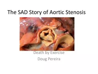
Symptoms • Asymptomatic (long latency period) • Decrease in exercise tolerance • Fatigue • Dyspnea • Angina • Syncope • Heart Failure
Signs (Physical Exam) • Loud, late-peaking systolic murmur • Radiation to the carotids • Single or paradoxically split S2 • Delayed and Diminished carotid upstroke (Pulsus parvus et tardus) • Low output states may affect intensity of the murmur and carotid upstroke • Associated increase in GI bleeding from angiodysplasia
EKG and CXR • Left ventricular hypertrophy (85 percent of patients) • Left atrial abnormality (80 percent) • Rounding of the left ventricular border with dilatation of the ascending aorta
Echocardiography • Qualitative • 2D imaging of valve anatomy (number of leaflets) • Calcification/thickening, valve opening • Left ventricular function • Chamber size, wall thickness • Quantitative • Peak aortic valve velocity • Mean and maximum pressure gradients • Aortic valve area (continuity equation) • Ratio of outflow tract to aortic jet velocity
Quantitative Doppler Assessment • Dedicated non-imaging transducer • Maximum pressure gradient using the simplified Bernoulli equation ∆Pmax = 4(Vmax)² • Mean pressure gradient obtained from planimetry of the Doppler envelope; computer integrates velocity data to provide mean value • Mean pressure gradient can also be estimated from published regression equations (for native valve AS): ∆Pmean = (∆Pmax/1.45) + 2mmHg
Quantitative Assessment • Pressure gradients are dependent on volume flow rates in addition to valve narrowing • Differences in stroke volume can lead to inaccurate assessment of stenosis severity • Coexisting AI will have a high transaortic pressure gradient • Patients with LV systolic dysfunction or coexisting MR may have low transaortic pressure gradient
Quality Control • Angle θ should be as close to zero degrees as possible, significant errors in measurement when greater than 20 degrees • Mistaken identity (MR jet) • Locating highest velocity signals • Recognize differences in reported data (peak instantaneous gradient vs peak-to-peak gradient)
Continuity Equation Valve Area • Use principle of continuity of flow to calculate valve area; not affected by changes in SV (as when increased in AI and decreased SV with LV dysfunction) • LV outflow tract diameter • Measured on 2D PSLA midsystolic image • Presume a circular shape • Measure diameter (D), one-half of measured is radius (r) • Area = лr² • LVOT VTI • Pulsed Doppler from apical approach, position sample just proximal to the stenotic valve • Aortic jet VTI • Continuous-wave Doppler from the window that yields the highest signal • For clinical use, may simplify by substituting maximum velocities for velocity-time integrals (Otto)
Velocity Ratio • Define “normal” valve area for the individual as the cross-sectional area of the outflow tract • The increase in velocity from outflow tract to aortic jet reflects stenosis severity regardless of size • Actual AVA / “normal” AVA ≈ (V lvot / V Ao) • Ratio of 1 indicates little obstruction, 0.5 half normal and 0.25 one-fourth normal value.
Low gradient with LV dysfunction • At very low flow rates, valve opening may be inhibited leading to an underestimation of aortic valve area • May be difficult to distinguish severe AS with low-pressure gradient due to low SV from mild-moderate stenosis with reduced Ao valve opening due to low flow. • Dobutamine echocardiography can help distinguish • Infuse 5mcg/kg/min in 5 mcg increments every 3 minutes until LVOT velocity or TVI reaches a normal value (usually at 15-20 mcg/kg/min) • True severe AS will have a fixed valve area • Consider severe if valve area < 1cm2 or > 30mmHG gradient with dobutamine • Also used to assess inotropic reserve (increase in SV > 20% with dobutamine infusion) • Class IIa (B)
Natural History of Progression • Aortic jet velocity: 0.3 m per second per year • Increase in mean gradient pressure of 7mmHg per year • Decrease in valve area of 0.1cm² per year (JACC Vol 48 (3): e1-148.)
Management • Presence of absence of symptoms • Severity of stenosis • Status of the left ventricle • Presence of comorbidities
Follow-up Exams • An optimal schedule for repeated medical examinations has not been defined. • An annual history and physical examination on patients with asymptomatic AS of any degree is reasonable. • Annual echocardiogram for severe AS (Class I, LOE: B) • An essential component of each visit is patient education about the expected disease course and symptoms. • Because the rate of progression varies considerably, clinicians often perform an annual echocardiogram every year which may be appropriate. • In patients with moderate AS, serial studies performed every 1 to 2 years are satisfactory. Mild AS, serial studies can be performed every 3 to 5 years (Class I, LOE: B). • Echocardiograms should be performed more frequently if there is a change in signs or symptoms.
Cardiac Catheterization • Class I • 1. Coronary angiography is recommended before AVR in patients with AS at risk for CAD (Level of Evidence: B) • 2. Cardiac catheterization for hemodynamic measurements is recommended for assessment of severity of AS in symptomatic patients when noninvasive tests are inconclusive or when there is a discrepancy between noninvasive tests and clinical findings regarding severity of AS. (Level of Evidence: C) • 3. Coronary angiography is recommended before AVR in patients with AS for whom a pulmonary autograft (Ross procedure) is contemplated and if the origin of the coronary arteries was not identified by noninvasive technique. (Level of Evidence: C)