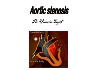
Aortic Stenosis
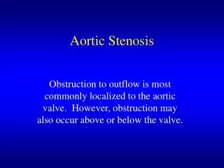
Aortic Stenosis. Obstruction to outflow is most commonly localized to the aortic valve. However, obstruction may also occur above or below the valve. Netter. Aortic Stenosis Etiology. Congenital Unicuspid produce severe obstruction in infancy and is fatal Bicuspid Valves

Aortic Stenosis
E N D
Presentation Transcript
Aortic Stenosis Obstruction to outflow is most commonly localized to the aortic valve. However, obstruction may also occur above or below the valve.
Aortic StenosisEtiology • Congenital • Unicuspid produce severe obstruction in infancy and is fatal • Bicuspid Valves • Occurs in 2% of the population and is the most common congenital cardiac defect in the adult • Presents with stenosis earlier in life • Abnormal architecture leads to turbulent flow and fibrosis
Normal and Congenital Valves Tricuspid Valve Unicuspid Valve Bicuspid Valve
Aortic StenosisEtiology • Acquired • Rheumatic • Results from adhesion and fusion of the commissures and cusps leading to retraction and stiffening of the free borders with calcific nodules • The valve is often regurgitant as well, and is often accompanied by evidence of MV involvement • Degenerative (Senile) • The cusps are immobilized by a deposit of calcium along the flexion lines in their bases
Rheumatic and Calcified Valves Calcific Bicuspid Valve Rheumatic Valve Calcific Tricuspid Valve
Mixed Valves Congenital Bicuspid Valve affected by Rheumatic Disease and Calcification Tricuspid Valve with Rheumatic Disease creating a functional bicuspid valve, and calcification
Aortic StenosisClues to diagnosis Aortic Stenosis Aortic Regurgitation Isolated AS or MV involvement with calcification Rheumatic under 70 yrs. Old over 70 yrs. Old bicuspid valve senile degeneration
Aortic StenosisPathophysiology Outflow obstruction Outflow Resistance Concentric Hypertrophy Maintain CO & SV LV Compliance Diastolic Pressure Enhanced LA contraction Maintain LV filling S4
Aortic StenosisClinical Manifestations • History • Angina • Occurs in 2/3 of patients with critical AS • Half of the patients have normal coronaries • Results from increased oxygen demand by a hypertrophied myocardium and decreased oxygen delivery secondary to compression of the vessels • Average survival is 5 years
Aortic StenosisClinical Manifestations • History • Syncope • Due to reduced cerebral perfusion • May be orthostatic, exertional, medication related (nitrates, diuretics, etc.), or due to arrhythmias • Average survival is 3 years
Aortic StenosisClinical Manifestations • History • Heart Failure • Manifest as orthopnea, dyspnea, PND, pulmonary edema • Average survival is 1 – 2 years
Aortic StenosisClinical Manifestations • Physical Examination • Venous System • Venous pulse configuration and pressure are unremarkable in well compensated AS • An increased A wave may occur as a result of decreased RV compliance secondary to LVH (Bernheim effect)
Aortic StenosisClinical Manifestations • Physical Examination • Carotid Arterial Pulse • The classic arterial pulse is called pulsus parvus et tardus (slow and late) • Precordium • The apical impulse has a sustained lift • There is little or no leftward displacement of the PMI
Aortic StenosisClinical Manifestations • Physical Examination • Auscultation • S1 – usually normal, may be soft if CHF present • S2 – the intensity of A2 decreases as the valve stiffens • S2 splitting – with prolongation of LV ejection time A2 will occur later than P2 and cause paradoxical splitting of S2
Aortic StenosisParadoxical Splitting P A A P Inspiration Expiration
Aortic StenosisClinical Manifestations • Physical Examination • S3 – usually not a normal finding in aortic stenosis, it’s presence suggests LV dysfunction • S4 – is usually present and suggests LV hypertrophy and decreased LV compliance • Ejection click occurs when the leaflets abruptly halt after maximal upward excursion and imply a mobile valve. It disappears as the valve becomes severely calcified.
Murmur of Aortic Stenosis Heard best at the 2nd RICS radiating to the carotids, sometimes throughout the precordium. S 2 S4 S 1
Aortic Stenosis Severity Mild Moderate Severe
tee Normal Tricuspid Valve Calcified Valvular Stenosis
Aortic StenosisNatural History • Symptomatic Patients • Angina = 5 year • Syncope = 3 year • CHF = 1-2 years • Asymptomatic Patients • 4% risk of sudden death
Aortic StenosisMedical Management • All patients should follow SBE prophylaxis guidelines • Avoid vigorous exercise • Use nitrates and diuretics with caution • Asymptomatic patients should report the onset of any symptoms promptly