Download
1 / 61
620 likes | 956 Vues
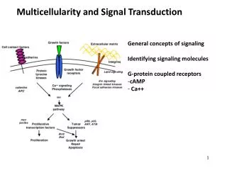
Diseases assotiated with abnomal signal transduction and cellular proliferation and apoptosis . Basal concept. Cell proliferation Cell entered the cell cycle sequentially under the accurate regulation. Significance of cell proliferation 1. Cell renewing

E N D
Diseases assotiated with abnomal signal transduction and cellular proliferation and apoptosis
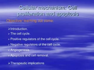
Basal concept • Cell proliferation Cell entered the cell cycle sequentially under the accurate regulation. Significance of cell proliferation 1. Cell renewing 2. Providing resource for cell differentiation 3. Supplement of dead cells
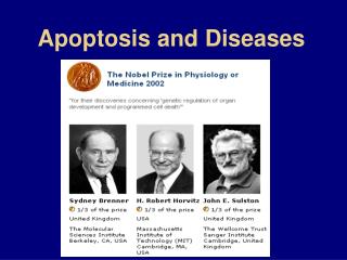
Cell apoptosis Apoptosis is a form of programmed cell death in multicellular organisms triggered by external and internal signals under some physio- or pathological conditions. Significance of apoptosis 1. Maintaining homeostasis of the body 2. Facilitating development of organs 3. Removing the damaged cell
Article 1Regulation of cell proliferation and apoptosis One、Cell proliferation and apoptosis are regulated by genes. Two、Regulation is implemented by gene products of oncogenes and anti-oncogenes.
One、Gene regulation of cell proliferation and apoptosis (1) cell proliferation and its regulation 1.cell cycle definition: a period from the end of one division to the beginning of next division of a proliferative cell. (G0—G1—S—G2—M) 2.Check point of cell cycle G0/G1、G1/S、G2/M 3. Major cell cycle regulatory proteins Cyclin, subtype: A-I, K、T) Cyclin-dependent kinase, CDK1-10) CDK inhibitor, CDKI) INK4 family: P15/16/18/19 CIP/KIP family:P21/27/57
cyclin cyclin cyclin Activity form cyclin cyclin Role of CDK in cell cycle
(2)Proteins involved in cell apoptosis 1.Caspase family 2.Bcl-2family anti-apoptotic: Bcl-2 pro-apoptotic:Bax、Bak/Bid、Bad, et al. 3. Tumor necrosis factor receptor,TNFR Fas(CD95)、TNF-R1、DR4、DR5
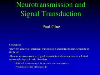
Two、 Regulation of onco-gene and anti-oncogene by signal transduction Normal cells:Reproduce themselves exactly, stop reproducing at the right time, stick together in the right place, self destruct if they are damaged or become specialized or mature. Onco-gene and anti-oncogene:Main genes of regulating cell proliferation and differentiation Cancer:Is a class of diseases in which a group of cells display uncontrolled growth, invasion, and sometimes metastasis resulted from the disorder of cell proliferation, differentiation, aging and death. Mechanisms:mutations of onco- and anti-oncogenes caused by mutagens and unstability of genes
DNA mutation causes abnormal cell proliferation and apoptosis Normal cell Successful DNA repairing DNA damage factor DNA damage Genetic faults DNA repairing failure DNA repair gene inactivation Somatic cell gene mutation Oncogene activation Tumor suppressor inactivation Change of appoptosis regulatory gene expression Quantitative and qualitative changes of gene products Unbalance in cell proliferation and apoptosis Degenerative disease in nervous system (over apoptosis) Autoimmune disease (insufficient apoptosis) tumor
Article 2Oncogene, anti-oncogene and signal transduction mediated by onco- and anti-oncogenes • Oncogene • Signal transduction associated with cell growth • Anti-oncogene (tumor suppressor gene )
One、oncogene, onc Basal concept • These genes code for proteins that are capable of stimulating cell growth and division. • In normal tissues and organisms, such growth-stimulating proteins are regulated, so that growth is appropriately limited. • However, changes/mutation in these genes may result in loss of growth regulation, leading to uncontrolled cell proliferation and tumor development. • These changed genes are known as oncogenes, because they induce the oncogenic state — cancer. • Oncogenes are dominant, because a change/mutation of only one allele of that gene can lead to tumor formation.
(1)Oncogene 1. Virus oncogene, v-onc • These genes are in viruses, may lead to uncontrolled cell proliferation and tumor development. • Virus oncogenes arehomolog with that corresponding cellular oncogenes. No intron Mutation pattern
Retrovirus (Chronic transforming retrovirus — wild type) RNA virus: containing gene coding for transcriptase • Basic structure and function of retrovirus Genome: Two copies of double strand RNA, 3~9Kb • Three structural genes (5’-gag--pol--env--3’) : gag--- core protein pol---transcriptase env---coat protein • Long terminal repeats, LTR promoter enhancer adding signal sequence of polyA
General structure of retrovirus LTR LTR
env gag pol
provirus Infectious course of retrovirus
Provirus: A provirus is a DNA mediate with LTR at both ends that has integrated itself into the DNA of a host cell. Provirus→expression→packaged into new viral article→budding outside cell surface • Resource of v-onc Definite DNA sequence captured from host cell example: src ALV(no v-src)--------------→ASV(containing v-src) ( avian sarcoma virus, ASV )
Obtaining of v-onc Provirus DNA Cell genome DNA Integrating DNA Transcripting, splicing Virus DNA Packaged into virus article
Acute transforming retrovirus • Structure features: ① Having different types of defects in structural genes, replication defect, non-infective virus ② Inserting definite sequence-oncogene ③ Only containing one sort of oncogene • Infection features: ① Short latent period ② Having ability to transforming the corresponding target cells in vitro into malignant cells • Exceptions : ① Avian sarcoma virus, ASV Oncogene locates in downstream of structural gene and does not breakdown structural gene of virus. ② Avian erythroblastosis virus, AEV Containing two sorts of oncogenes: erb-A and erb-B
2.cellular oncogene, C-onc • Real copy of v-onc in eukaryotic cell.An cellular oncogene is an activated form of pro-oncogene. A proto-oncogene is a normal gene that can become an oncogene due to mutations or increased expression. Proto-oncogenes code for proteins that help to regulate cell growth and differentiation—house-keepinggenes. Uponactivation, a proto-oncogene (or its product) becomes a tumor-inducing agent, an oncogene. • Proto-onc: In normal cells, c-onc exists in non-activated form.
Features of c-onc • (1) Structral features:with the general structure of eukaryotic genes; Different creatures: introns--with greater difference exons --conservative sequence • (2) Widespread in living nature; • (3) Highly conservative in gene sequence; • (4) Executing function by products of protein; • (5)Uponactivation ( mutations or increased expression ), becomes a tumor-inducing agent, an oncogene.
Differences between v-onc and c-onc: • V-onc often loses some sequences at both ends • No introns in v-onc • V-onc is the mutated form of c-onc
Classification of c-onc • There are several systems for classifying oncogenes,but there is not yet a widely accepted standard. They are sometimes grouped both spatially (moving from outside the cell inwards) and chronologically (parallelling the "normal" process of signal transduction). • Pro-onc mains codes for key molecules involved in signal transduction and gene transcription.
1.Growth factors • sisgene:Homologous with B chain of PDGF • int-2, hst:Homologous with fibroblast growth factor ( FGF) 2. Growth factor receptors • C-erb-B:EGFR • C-fms:Colony stimulating factor receptor(CSF-1) • bit:PDGFR • ros:insulin receptor • mas:angiotonin (AGT)receptor or αreceptor TPK
3. Non-receptor tyrosine protein kinases • src • abl 4.GTP binding protein • H-ras、K-ras、N-ras ( 21KD- small G protein ) 5. Intranuclear DNA binding protein • Fos-Jun(transcription factor AP-1)→TRE(TPA reaction element) • CREB-Jun→CRE • C-myc(basic amio acid )→single/double strand DNA • Rel(NF-κB associated protein)
(2)Activation mechanisms of pro-onc 1.Insertion of regulatory sequence ( promoter, enhancer) ① ALV with v-onc induces B cell lymphoma ② Experiment of Leder group( trans-oncogene animal model) LTR of mouse mammary tumor virus ( MTV ) +C-myc(MTV/myc) 2. Gene mutation Gene mutation within key modulin of cell carcinoma of bladder : T24 cell C-rasH T35 --- Val 12 normal: G35 --- Gly 12 3.Gene rearrangement • Burkitt lymphoma:8q24(c-myc) 14q32 close to immune globulin There is a strong enhancer in heavy chain region of immune globulin, and causes overexpression of C-myc . • Philadelphia chromosome ( balanced translocation ):9q34 22q11 C-abl interchanging with bcr
Gene translocation —Burkitt lymphoma TCR:T cell receptor
Chromosome 22-bcr gene Chromosome 9-c-abl gene Breakpoint in ALL Breakpoint in CML(bcr=5.8kb) Breakpoint in AML and CML Gene translocation — Philadelphia chromosome 160KD of Bcr protein 140KD of Abl protein fusion gene of bcr-abl in ALL 18.5KD of fusion protein fusion gene of bcr-abl in CML 210KD of fusion protein ALL: Acute lymphoblastic leukemia ; CML: Chronic myelogenous leukemia
4. Gene amplification ① Homogeneously staining regions, HSR Homogeneously staining regions except normal staining regions ② Double minutes, DM Punctiform pseudochromosome 5.Gene couple Under certain conditions, some pro-oncs are sequentially activated due to activation of other pro-oncs. Products of oncogene promoting immortalization of cell locate in nucleus. (C-myc) Products of oncogene promoting proliferation of cell locate in cytoplasm. (C-ras) 6.Elimination of intergenic suppression Special fragment regulating gene expression ① No.1 exon of C-myc gene dose not encode protein, and may have inhibitory action ,if loss, causes activation of c-myc. ② Methylation of DNA increases stability of double helix.
(3)Function of oncogene • Immortalization of cell Blocking cell differentiation,retrieving cell apoptosis, often locating in nucleus. • Transformation of cell Decreasing dependence of cell proliferation on growth factors,often locating in cytoplasm or cell membrane.
GRB2 SOS Raf MAPKK P P P P P MAPK fos、jun、myc Regulating gene expression (cyclinD) Regulating activity of other proteins Extracellular signal: EGF、PDGF and so on. Two、Main signal transduction pathway associated with growth Pate cancer、Breast cancer Cell membrane Ras- GTP Having mutation in 90% malignant tumor PI3K - Ovarian cancer (MAPKKK) PIP3 PTEN Akt (MEK) Anti-apoptosis、promoting proliferation Neuclus (ERK)
Three、Tumor suppressor gene • Concept A tumor suppressor gene, or anti-oncogene, is a gene that protects a cell from one step on the path to cancer. Tumor-suppressor genes, or more precisely, the proteins for which they code, either have a dampening or repressive effect on the regulation of the cell cycle or promote apoptosis, and sometimes do both. 1. Mostly are recessive genes and not easy to be discovered. 2. Theoretically, the number of oncogenes matches that of tumor suppressor genes. 3. The position of oncogene and tumor suppressor gene may be equally important.
Discovery of tumor suppressor gene: • Cognitive process:oncoprotein-oncogene-tumor suppressor gene 1. Analysis of cell genetics 70s,Kundson—Predisposing gene of retinal glioblastoma (Retinoblastoma,Rb)deletion —Existence of tumor suppressor genes 2. Experiment of cell hybridization 70s,Harris—Hybridoma experiment —Proving the exsitence of tumor suppressor gene
Cell fusion Normal cell Nontumorous hybrid cell Tumor cell Tumor cell1 Cell fusion Nontumorous hybrid cell Tumor cell2 Loss of some genes Normal cell Tumor cell Experiment of cell hybridization
p53– Predisposing gene of many kinds of tumors • P53 — So far, the most relevant gene with human tumor. • Mutation form 1. Complete loss of p53 gene 2. p53 gene mutation : ① Two allelic inactivation of p53 ② One allele for p53 gene inactivation ,molecules of p53 with mutation dimerise with wild-type p53 and prevent them from executing functions.
Acid region Core region Basic acid p53 1. Gene localization:17p13.1 2. Gene structure:11 exons,No.1 does not code for protein. 3. Protein structure:53kDa ( 7 domains,Th:20min ) -C’ N’- Homo-oligomerisation ( tetramerization ) domain:many modification sites associated with regulating DNA binding activity. Easy to be hydrolyzed by protease ;Phosphorylation site associated with protein stabibility. DNA binding region— Important functional position
p53 pathway stability Transcription activity Activation form
Replication failure P21gene Mechanism of Growth arrest induced by P53 Replication facor A Successful repair DNA damage P53 protein Cytocidal P53 protein P53 protein suppression P21protein Unwinding enzyme Holding the cell cycle at G1 phase
Retinoblastoma,Rb 1. Gene localization:13q14 2. Gene structure:27 exons. 3. Protein structure:105kDa(P105RB) 4. Protein function:DNA binding protein,binding E2F at phase G0andG1; Inhibiting transcription activity of E2F; Inhibiting expression of many oncoproteins; Regulating cell cycle. 5.Activity regulation: Phosphorylation decreasing its activity; Dephosphorylation activation. 6.Regulatory factor: Cyclin-CDK Oligomerisation region Binding region of E2F and viral oncoprotein DNA binding region
Rb protein E-2F Rb protein P Mechanism of action of Rb gene G0 ,G1phase DNA S phase E-2F DNA mRNA
Kundson :Tow-hit hypothesis An inherited, germ-line mutation in a tumor suppressor gene (Rb) would only cause cancer if another mutation event occurred later in the organisms’s life, inactivating the other allele of that tumor suppressor gene. • * It shows for the first time that disease of dominant heredity in family has recessive mutant tumor suppressors alleles. • * Mutation form of Rb: Gene deletion and gene mutation; Also associated with osteogenic sarcoma, prostatic carcinoma, small cell lung cancer and so on.