Methods
0.5 cm. MCF7. ER. cMyc. ER. cMyc. r1 app (ER+)=28.5±0.1, n=2 r1 app (ER-)=19.6. 3. 2. 1. 0. agonist. mild-agonist. In Vitro and In Vivo Molecular Imaging of the Estrogen Receptor using Novel ER -Targeted MRI Contrast Agents.

Methods
E N D
Presentation Transcript
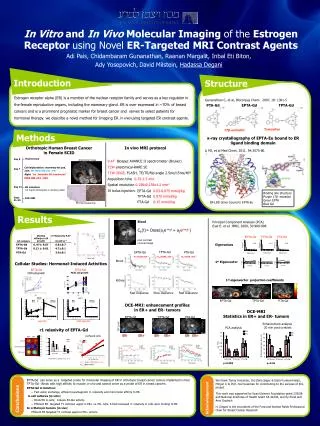
0.5 cm MCF7 ER cMyc ER cMyc r1app (ER+)=28.5±0.1, n=2 r1app(ER-)=19.6 3 2 1 0 agonist mild-agonist In Vitro and In Vivo Molecular Imaging of the Estrogen Receptor using Novel ER-Targeted MRI Contrast Agents AdiPais, Chidambaram Gunanathan, RaananMargalit, InbalEtiBiton,AdyYosepovich,David Milstein, HadassaDegani Introduction Structure TPTA-Gd PTA-Gd EPTA-Gd Estrogen receptor alpha (ER) is a member of the nuclear receptor family and serves as a key regulator in the female reproductive organs, including the mammary gland. ER is over expressed in ~70% of breast cancers and is a prominent prognostic marker for breast cancer and serves to select patients for hormonal therapy. we describe a novel method for Imaging ER in vivo using targeted ER contrast agents. Gunanathan C, et al,Bioconjug Chem. 2007, 18: 1361-5 * * PTA-Gd EPTA-Gd TPTA-Gd tamoxifen 17β-estradiol Tamoxifen 17β-estradiol Methods x-ray crystallography of EPTA-Eu bound to ER ligand binding domain Orthotopic Human Breast Cancerin Female SCID In vivo MRI protocol Li MJ, et al Med Chem. 2011, 54:3575-80. Day 0 Ovariectomy Anatomical image 9.4TBiospec AVANCE II spectrometer (Bruker) T2W anatomical-RARE SE T1W-3DGE, FLASH, TE/TR/flip angle 2.5ms/15ms/40º Acquisition time 0.75-1.5 min Spatial resolution 0.156x0.156x1.2mm3 IV bolus injection: Day 7 Cell implantation:mammary fat pad, Left:WT MDA-MB-231 : ER- Right:Tet Inducible ER transfected MDA-MB-231 : ER+ ER- ER+ Day 14 ER induction: 0.2 mg/ml tetracycline in drinking water EPTA-Gd0.03-0.075mmol/kg TPTA-Gd0.075 mmol/kg PTA-Gd0.15 mmol/kg Binding site structure Purple 17β- estradiol Green EPTA Blue Gd Days 21-35 DCE-MRI ER-LBD dimer bound to EPTA-Eu ER Immunostaining Results Blood Principal Component Analysis (PCA) Eyal E. et al. JMRI, 2009, 30:989-998 Cb(t)= Dose(a1e-m1t + a2e-m2t ) EPTA-Gd TPTA-Gd PTA-Gd post contrast coronal image TPTA-Gd EPTA-Gd PTA-Gd Eigenvalues m2=0.03 min-1 m2=0.003, min-1 m2=0.04, min-1 Blood Cellular Studies: Hormonal-Induced Activities 1st Eigenvector EPTA-Gd TPTA-Gd Kidney MCF7 cell growth T47D cell growth 1st eigenvector projection coefficients Fast clearance Slow clearance Fast clearance 0.5 cm DCE-MRI Statistics in ER+ and ER- tumors DCE-MRI: enhancement profiles in ER+ and ER- tumors EPTA-Gd TPTA-Gd PTA-Gd MCF7 Enhancement analysis 20 min post contrast EPTA-Gd TPTA-Gd PTA-Gd PCA analysis 20’ post ER- ER+ ER- ER+ ER- ER+ • r1 relaxivity of EPTA-Gd • perfused cells p<0.008 p<0.05 N=9 N=4 N=4 N=9 N=4 N=4 * Pval<0.035, paired t-test, n=9 • EPTA-Gd can serve as a targeted probe for molecular imaging of ER in orthotopic breast cancer tumors implanted in mice • TPTA-Gd - Binds with high affinity to muscle in vivo andcannot serve as a probe of ER in breast cancers. We thank Tamar Kreizman, Drs Dalia Seger & Edna Furman-Haran, MinjunLi & Prof. Joel Sussmanfor contributing to the success of this project This work was supported by Israel Science Foundation grant 235/08 and National Institutes of Health Grant CA 42238, and by Ernst and Anni Deutsch H. Degani is the incumbent of the Fred and Andrea Fallek Professorial Chair for Breast Cancer Research EPTA-Gd in Solution: • Fast water exchange, efficient paramagnetic r1 relaxivity and micromolar affinity to ER. in cell cultures (in vitro) • Binds ER in cells; induces E2-like activity. • Efficient ER targeted T1 contrast agent in ER+ vs. ER- cells, 5-fold increased r1 relaxivity in cells upon binding to ER. • In orthotopic tumors (in vivo) • Efficient ER targeted T1 contrast agent in ER+ tumors. Conclusions Acknowledgement