Methods
Biomarkers of cartilage degradation and synthesis relevant to knee osteoarthritis: Relationships with dynamic knee joint load and changes following exercise. Michael A. Hunt 1,2 , Courtney L. Pollock 1 , Virginia Byers Kraus 3 , Tore Saxne 4 , Sue Peters 1, Jolanda Cibere 2

Methods
E N D
Presentation Transcript
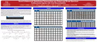
Biomarkers of cartilage degradation and synthesis relevant to knee osteoarthritis: Relationships with dynamic knee joint load and changes following exercise Michael A. Hunt1,2, Courtney L. Pollock1, Virginia Byers Kraus3, Tore Saxne4, Sue Peters1, Jolanda Cibere2 1Department of Physical Therapy: University of British Columbia Vancouver, Canada; 2Arthtritis Research Centre of Canada: Vancouver, Canada; 3Duke University Medical Center: Duke University Raleigh-Durham, USA; 4Department of Rheumatology: Lund University Lund, Sweden Introduction High knee joint loads during walking have been implicated in the pathogenesis of knee osteoarthritis (OA) [1]. The external knee adduction moment (KAM) during walking gait has received attention as a valid proxy of medial compartment knee joint load with clinical relevance to knee OA, including the ability to predict the risk of OA progression [2] resulting from cartilage degradation. Concurrently, the use of cartilage-derived biomarkers to examine direct processes of cartilage degradation and synthesis are increasingly being used to examine clinically relevant aspects of knee OA. Specifically, a number of OA biomarkers have been reported to be higher in those with more severe OA [3] and have shown an ability to predict OA progression [4]. However, very limited research has been conducted examining the influence of knee joint load on biomarker levels or changes in levels following treatment. Figure 1: The KAM impulse was calculated as the area under the KAM-time curve, an outcome suggested to be more indicative of knee load throughout the gait cycle. Figure 2: Scatter plot of KAM impulse vs. unadjusted uCTX-II values at baseline (n=17 participants). Note that higher KAM impulse values were associated with more cartilage degradation as quantified by log-transformed uCTX-II concentrations. Figure 3: Scatter plot of KAM impulse vs. unadjusted uCTX-II:sCPII values at baseline (n=17 participants). Note that higher KAM impulse values were associated with higher log-transformed ratio values indicating more cartilage degradation (uCTX-II) compared to synthesis (sCPII). Results At baseline after adjusting for age, sex, and physical activity, KAM impulse was only significantly predictive of uCTX-II levels (p = 0.02) and the ratio uCTX-II:sCPII (p < 0.01). (Figures 2,3) Following the intervention, those in the exercise group exhibited significantly greater reductions in sCOMP (p = 0.04) and a trend towards greater reductions in uCTX-II (p = 0.10) compared to the no exercise group. (Table 1) Table 1: Mean (sd) values for log-transformed, unadjusted biomarker levels and ratios of degradation to synthesis (sCPII) at baseline and follow-up for each group. Group comparisons (exercise – no exercise) denote the difference in mean change (95% CIs) for each log-transformed biomarker and ratio after adjusting for age, sex, and physical activity (PASE). • Objectives • To examine the relationships between dynamic knee joint load and cartilage biomarker levels in people with knee OA • To compare changes in cartilage biomarker concentrations following an exercise intervention aimed at cartilage offloading in people with knee OA. Conclusions This research provides possible evidence of a direct relationship between joint load and production of uCTX-II and sheds new light on the validity of this biomarker relevant to knee OA. Further, exercises designed to strengthen muscles and offload the medial knee joint can significantly reduce the circulating levels of OA degradation biomarkers (sCOMP, uCTX-II). Taken together, this research provides new information on the clinical utility of OA biomarkers and new outcomes for shorter term OA interventions such as exercise. ** note that negative log-transformed values indicate that the absolute ratio was less than 1.0, with greater negative values indicating a smaller ratio of the degradation biomarker to the synthesis biomarker sCPII. Thus, improvements in the ratio of degradation to synthesis would be reflected in larger negative values at follow-up. • Methods • 17 participants (8 male, 9 female; mean (sd) 66.1 (11.3) years of age with radiographically-confirmed knee OA were assessed on two separate occasions, eleven weeks apart. The concentrations of OA-relevant biomarkers measured from serum and urine as well as the magnitude dynamic knee joint load during 3-D gait analysis (KAM) were quantified at each testing session. Physical activity was quantified using the Physical Activity Scale for the Elderly (PASE). • 3-D motion analysis was conducted using 8 high-speed digital cameras synchronized with two floor-mounted force platforms. 22 reflective markers (Helen Hayes marker set) were used to obtain kinematic data as participants completed five walking trials at a self-selected gait speed . Inverse dynamics were used to calculate the KAM for each trial and the KAM impulse (area under the KAM-time curve) was calculated and averaged across the five trials at each session (Figure 1). • From the serum and urine samples, the following biomarkers were measured using commercially available assays: urinary C-telopeptide of type II collagen (uCTX-II) and type II collagen cleavage neoepitope (uC2C), both normalized for creatinine; serum cartilage oligomeric matrix protein (sCOMP); serum hyaluronic acid (sHA); and serum C-propeptide of type II procollagen (sCPII). • Following baseline testing, participants were randomly assigned to one of two groups: 1) exercise, or 2) no exercise. Those in the exercise group were provided with six exercises designed to strengthen the quadriceps, hamstrings, and hip abductor groups and improve lower limb dynamic alignment. Exercises were completed at home four days per week and participants met with the study physiotherapist five times over ten weeks to ensure proper performance and safe progression of resistance. Those in the no exercise group were not provided with any additional treatment and were instructed to maintain their usual care. • Regression models were used to examine the contribution of joint load on biomarker concentrations while adjusting for age, sex, and physical activity. Regression modeling was also to compare changes in each biomarker level between groups. • References • Andriacchi and Mundermann. CurrOpin Rheum 2006;18:514-518. • Miyazaki et al. Ann Rheum Dis2002;61:617-622. • Cibere et al. Arthritis Rheum 2009;60:1372-1380. • Reijman et al. Arthritis Rheum 2004;50:2471-2478 Sources of funding: