Download
1 / 12
200 likes | 800 Vues
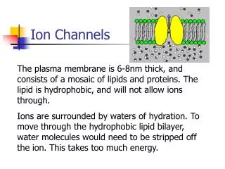
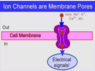
Types of Ion Channels Leak channels some are always open (open and close randomly) e.g., K + leak channels cause nerve and muscle cell membranes to be more permeable to K + than to Na + .

E N D
Types of Ion Channels • Leak channels • some are always open (open and close randomly) • e.g., K+ leak channels cause nerve and muscle cell membranes to be more permeable to K+ than to Na+. • Gated channels (Even though the behavior of an individual channel may appear to be random, the stimulus significantly changes the probability of a population of channels being open or closed.) • voltage-gated channels • open/close due to changes in membrane potential • e.g., voltage-gated channels for Na+, K+, and Ca++ • ligand-gated channels (“chemically-gated”) • open/close due to the binding (ligation) of a chemical signal (ligand) • e.g., The nicotinic cholinergic receptor opens when acetylcholine (ACh) binds to it. • mechanically-gated channels • open/close due to a mechanical stimulus • e.g., sensory receptors that respond to pressure or stretch
Examples of Gated Channels e.g., voltage-gated K+ channel e.g., nicotinic cholinergic receptor Fig. 11-21 Alberts et al., Molecular Biology of the Cell
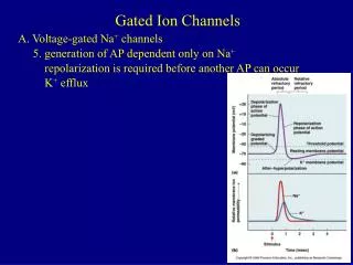
Fig. 14-2 Katzung, Pharmacology Voltage-gated Na+ Channel three states: resting, activated, and inactivated resting activation gate (m) closed, inactivation (h) gate open • activated • activation gate open, inactivation gate open • stimulus for opening: depolarization • time to start opening: ~ 0.1 msec • inactivated • activation gate open, inactivation gate closed • stimulus for closing: depolarization • time to start closing: ~ 0.2 msec recovery: When the membrane returns to the resting membrane potential, the Na+ channels return to the resting state.
Voltage-gated K+ Channel two states: closed and open Fig. 12.14 plus Fig.11-21, Alberts • open • stimulus for opening: depolarization • time to start opening: ~ 0.2 msec • closed • during resting state • inactivated • In some neurons (e.g., rapidly firing neurons) the K+ channel also has an inactivated state. Alberts et al., Molecular Biology of the Cell
Voltage-gated Na+ and K+ Channelsone stimulus, three responses /activated Panel 11-3, Alberts et al., Molecular Biology of the Cell
Fig. 12.14 - altered with channel cartoons from Katzung and Alberts Action Potentials changes in the channels Stimulus t = 0 0.1 ms [positive feedback] 0.2-0.3 ms
Reminder: Since depolarization, repolarization and after-hyperpolarization are due to the diffusion of Na+ and K+ through voltage-gated Na+ and K+ channels, the membrane potential cannot go higher than ENa (e.g., +60 mV) or lower than EK (e.g., –90 mV). Moffett, Moffett and Schauf, Human Physiology
Role of ATP • ATP is not directly needed for the production of an action potential. • Very few ions cross the membrane. So few that depletion of the sodium and potassium gradients takes hundreds of thousands of action potentials. • Over the long term, however, ATP energy is required to empower the Na+ pump to restore the transmembrane sodium and potassium gradients. • “battery charger” function of the sodium pump
Refractory Periods Moffett, Moffett and Schauf, Human Physiology Fig. 12.15 • Absolute refractory period: No stimulus can cause an action potential. • Na+ channels become inactivated and cannot open. • Relative refractory period: A greater than normal stimulus is required in order to trigger an action potential. • The distance to threshold is greater: 1) the threshold is higher • and 2) the membrane is hyperpolarized (more negative).
Local Anesthetics Local anesthetics of the “-caine” family (e.g., novocaine, benzocaine, lidocaine) block the Na+ channel at its intracellular end. Therefore, their effect is to put the nerve cell membrane into an absolute refractory period. anesthetic Fig. 14.2 Katzung, Pharmacology
Propagation of an Action Potential • It is possible for an electrical signal to travel along an axon or a muscle cell in either direction. • Nonetheless, in neurons the signal usually travels in only one direction. • Direction of movement is determined by what area is excited first.
Propagation of an Action Potential cell body and dendrites: “receiving end” axon terminals: “sending end” • One end of the cell is specialized to receive signals. Action potentials start at the part of the axon closest to the receiving end. • “Active” depolarization in one place causes depolarization-to-threshold “downstream.” • “Upstream” is refractory. • The “distal” end of the cell is specialized for sending signals to the next cell. A B C Fig. 12.16