Download
1 / 55
770 likes | 1.46k Vues
Cardiac Wellness Institute of Calgary. Pathophysiology and Risk Factors of Coronary Artery Disease. Updated May 2010. Overview. Cardiovascular Diseases Atherogenesis and response to Injury (Endothelial Dysfunction) Manifestations and Diagnosis of CAD Treatment of CAD

E N D
Cardiac Wellness Institute of Calgary Pathophysiology and Risk Factors of Coronary Artery Disease Updated May 2010
Overview • Cardiovascular Diseases • Atherogenesis and response to Injury (Endothelial Dysfunction) • Manifestations and Diagnosis of CAD • Treatment of CAD • Risk Factors Contributing to CAD • Modifiable vs Non-modifiable
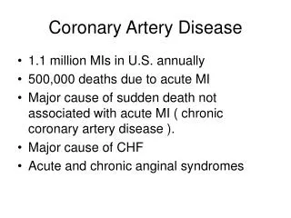
Cardiovascular Diseases • Arteriosclerosis – loss of elasticity of the arteries; thickening and hardening of artery walls. • Atherosclerosis – process where fatty material is deposited along walls of arteries. This material thickens, hardens, and can eventually block the artery. Atherosclerosis is just one type of Arteriosclerosis. • Our understanding of the development and progression of atherosclerosis (atherogenesis) is still incomplete
Vascular Anatomy • Endothelium: barrier between blood and arterial wall • 3 layers in arterial wall: • Tunica Intima - connective tissue; where lesions form • Tunica Media - smooth muscle • advanced atherosclerosis characterized by proliferation of smooth muscle cells here • Tunica Adventitia - connective tissue; highly vascularized to provide nutrients
Regulates vasomotion Regulates thrombosis Regulates transport of substances to and from vascular space Regulates growth and apoptosis of vascular wall Regulates LDL oxidation Inadequate vasodilation Prothrombotic Altered permeability Increased secretion of growth factors Increased oxidation of LDL Endothelial Function Endothelial Dysfunction
Atherogenesis Response to Injury • Arterial Injury • Can result from smoke, hypertension, cholesterol, glycated substances, vasoconstriction, homocysteine or infectious agents • Normal endothelial function is not repaired by inherent mechanisms • Endothelial Dysfunction and Inflammatory Response • Arterial homoeostasis is altered by injury, results in inflammatory response • Increased adhesiveness endothelial cells lose selective permeability
Atherogenesis • Platelet aggregation • Platelets adhere to damaged endothelium and form small blood clots on vessel wall (mural thrombi) • Release growth factors and vasoconstrictor substances • Can cause obstruction to blood flow
Atherogenesis • LDL oxidation • Excess oxidized LDL particles accumulate in arterial wall, attracting monocytes and other cells into intima • Monocytes mature into macrophages and cause proliferation of smooth muscle cells and promote uptake of more lipids, particularly LDL • These cells move from the media to the intima, becoming foam cells, producing fatty streaks or lesions • Continued release of vasoactive substances and growth factors
Atherogenesis • Foam cells • Release cholesterol into extracellular space • Fatty streaks • Earliest visually detectable lesion of atherosclerosis • As the process continues, smooth muscle cells accumulate in the intima and form a fibrous plaque
Response to Injury • Fibromuscular plaque • With continued accumulation, lesion progresses in size and appearance to Fibromuscular plaque with an Atheroma (cholesterol core) • Remodeling • Outward growth of artery & increased lumen size • Lumen size increases to compensate for atherosclerotic plaque • If plaque bulk continues to increase, lumen diameter is decreased and blood flow obstruction occurs
Atherogenesis • Plaque rupture, thrombus formation, incorporation • Layered appearance to lesion and increased plaque progression • Rupture may result from local stress or chemical factors and exposes contents or lesion to blood • Plaques that are most vulnerable to rupture typically have a large lipid core, thinned fibrous cap, and outward remodeling of arterial wall • Advanced Atherosclerotic Plaque
Progression of Atherosclerosis Coronary artery at lesion-prone location Type II (Lesion) Type III (Preatheroma) Adaptive thickening (smooth muscle) Small pools of extracellular lipid Macrophage foam cells Type VI (Complicated lesion) Type IV (Atheroma) Type V (Fibroatheroma) Thrombus fissure & hemtoma Core of extracellular lipid Fibrous thickening Adapted from Stary in Fuster et al (eds). Atherosclerosis and Coronary Artery Disease 1996.
Atherogenesis • Does not occur in a predictable linear pattern • Some lesions develop slowly and are stable for long periods of time, others develop quickly • Partial regression of fatty, soft lesions is possible with aggressive risk reduction • Endothelial dysfunction can be reversed • Exercise, dietary fat intake control, decreasing stress, maintaining optimal blood pressure and blood glucose levels
Manifestations of Atherosclerosis • The Heart • Myocardial Ischemia • Angina • Myocardial Infarction • Brain • Legs
Manifestations of Atherosclerosis Myocardial Ischemia – ischemic cascade • LV stiffening & decreased diastolic filling (diastolic dysfunction) • Impaired LV systolic emptying • ECG changes associated with altered repolarization • Angina Pectoris – transient, referred cardiac pain resulting from ischemia
Manifestations of Atherosclerosis Angina – Types: • Silent ischemia:no pain • Anginal Equivalent:shortness of breath, diaphoresis etc. • Typical Angina:occurs with exertion, emotions & relieved with rest or NTG • Atypical Angina:similar symptoms, but no exertion etc • Stable Angina:reproducible, predictable • Unstable Angina:new onset, increased freq, intensity, duration, or occurs at rest
Manifestations of Atherosclerosis Myocardial Infarction • Diagnosis: 2 of 3 criteria: 1) Chest pain > 30 minutes 2) ECG – Q waves / ST segment elevation/ T wave inversion 3) Cardiac enzymes: • Creatine phosphokinase (CK) Normal = 0-195 • Troponin T – Normal < 0.03
Manifestations of Atherosclerosis Myocardial Infarction • Signs & Symptoms: • Angina, GI upset, Dyspnea, Diaphoresis, Syncope • Treatment: • Relieve symptoms (nitroglycerin, painkillers) • Reperfusion
Manifestations of Atherosclerosis Myocardial Infarction • STEMI vs. NSTEMI: • ST Elevation MI – ST elevation of 1 mm or more in contiguous leads or new LBBB • Non-ST Elevation MI – ST depression or T wave inversion lasting greater than or equal to 24 hours
Manifestation of Atherosclerosis • Brain • Transient ischemic attack (TIA) • Cerebrovascular accident (stroke) • Legs • Intermittent claudication
Graded Exercise Test (GXT) Used to assess... • Ischemia • ST segment changes • Arrhythmia • Functional Capacity • MET’s • Efficacy of medical or surgical intervention
Myocardial Perfusion Imaging (Thallium scan) Used to assess... • Ischemia • Ventricular Function • Ejection Fraction • Myocardial Viability • Reversible vs non-reversible
Echocardiography Used to assess... • Myocardial Structures • MR, TR, AR • Ventricular Function • EF • Wall motion abnormalities • Effusions • Thrombus • Ischemia
Cardiac Angiography Used to assess... • Coronary arteries • Pressures within cardiac chambers • Valve function • Ventricular function
Risk Factor Modification Treat to Target
Lower the Demand Beta Blockers Decrease contractility Decrease heart rate Nitrates Decrease preload Calcium Channel Blockers Decrease preload Increase the Supply Beta Blockers Increase diastole Nitrates Increase collateral circulation Calcium Channel Blockers Decrease vascular resistance Medical ManagementBalancing the Supply and Demand Equation
Percutaneous Coronary Intervention (PCI) Indications for Angioplasty (+/- stenting) • Electively for chronic stable angina • Urgently for unstable angina • Emergently for myocardial infarction • 1 or 2 vessel disease • NEVER for left main disease
Coronary Artery Bypass Graft Surgery (CABG) Indications • Left main disease > 50 % • Proximal 3 vessel disease • Multivessel disease with left ventricular dysfunction • Lifestyle limiting angina unresponsive to medical therapy or PCI
Risk Factors for Coronary Artery Disease Non-modifiable and Modifiable
Non-Modifiable Risk Factors • Family History • Twice the risk of MI if one first-degree relative with MI • Triple the risk of MI if 2+ first-degree relatives with MI • Risk is strongest if MI occurred at age 55 or less • Advancing Age • Risk of CAD Increases as we get older • Gender • Men are at risk at an earlier age than women • Women’s risk of heart disease increases after menopause and soon equals men’s
Modifiable Risk Factors • Tobacco Smoking • Dyslipidemia • Hypertension • Obesity • Sedentary Lifestyle • Diabetes • Emerging Risk Factors
Tobacco Smoking • The MOST preventable risk factor • Smokers have 2 to 5 times the risk of CAD as nonsmokers • Risk factor if one is currently smoking, has quit within the past 6 months, or has exposure to environmental tobacco smoke
Tobacco Smoking • Increase workload to heart • Increased HR and BP • Endothelial dysfunction • Increased vasoconstriction • Decreased HDL • Increased LDL and Triglycerides • Increased LDL oxidation • Increased platelet aggregation • Decreased O2 carrying capacity of red blood cells
Dyslipidemia • 2 main types of lipids: • Cholesterol • Triglycerides (TGs) • Lipids are an essential component of healthy body functioning, including: • Structural component of cell walls • Hormones • Energy source
Dyslipidemia • Much research to support the link between abnormal serum lipid levels and CAD LDL = risk of CAD HDL = risk of CAD TGs = risk of CAD
Dyslipidemia • Abnormal lipid levels are known to be the basis of the atherosclerotic process • Endothelial Dysfunction • Elevated cholesterol levels • Reduce vasodilation • Increase thrombosis • Elevated triglyceride levels • Mechanism is unclear
Lipid Targets for CAD 2009 Canadian Cholesterol Guidelines Primary Targets: • LDL-C < 2.0mmol/L or 50% reduction • Alternate: Apolipoprotein B < 0.80 g/L Can J Cardiol 2009; 25(10): 567-579.
Lipid Targets for CAD 2009 Canadian Cholesterol Guidelines Secondary Targets:(once LDL cholesterol is at goal) • Total Cholestrol to High-Density Lipoprotein (HDL) cholesterol ratio less than 4.0 • Non HDL cholesterol < 3.5 mmol/L • Triglycerides < 1.7 mmol/L • Apolipoprotein B to apolipoprotein AI ratio < 0.8 • High-sensitivity C-reactive protein (CPR) < 2 mg/L Can J Cardiol 2009; 25(10): 567-579.
Hypertension • Primary risk factor for CAD • Hypertension is associated with three to four times increased risk for CAD, MI and CVA & PVD • Hypertension as a precursor or consequence of endothelial dysfunction? • Vasoconstriction (increases SBP) • Vascular wall injury • Increased platelet aggregation • myocardium • increased wall stress • increased myocardial O2 demand
Blood Pressure Targets ACSM Guidelines Optimal 120 / <80* Normal 120-129 / 80-84* High Normal 130-139 / 85-89* Hypertension >140 / >90* *All units in mmHg
Blood Pressure Targets 2010 Canadian Hypertension Guidelines • Non-Diabetics <140/90 mmHg • Diabetics or persons with chronic kidney disease <130/80 mmHg
Obesity • The risk for CVD is greater in person’s with central (android) obesity than those with peripheral (gynoid) obesity • Obesity is often associated with … • Diabetes • Hypertension • Dyslipidemia • Inactivity
Obesity • Body Mass Index (BMI) • Measured in Kg/m2 • ACSM BMI Targets Underweight <18.5 Normal 18.5-24.9 Overweight 25.0-29.9 Obese >30
Obesity • Waist Circumference • ACSM Waist Circumference Targets Men < 102 cm Women < 88 cm
Sedentary Lifestyle • Lower fitness level is associated with increased risk of CAD in men and women • The relative risk of CAD associated with physical inactivity is comparable to that observed for cigarette smoking, hypercholesterolemia and hypertension • Persons who are physically inactive after a heart attack have significantly high mortality rates than active individuals
Sedentary Lifestyle Physical activity reduces the risk of CAD through: • Improved balance between myocardial O2 supply and demand • Decreased platelet aggregation • Decreased susceptibility to malignant ventricular arrhythmias • Improved endothelial tone • Beneficial effect on other CAD risk factors (ie. diabetes, dyslipidemia, hypertension, obesity, stress)
Diabetes • People with diabetes have 2 to 7 times increased risk of developing CAD than people without diabetes • Mechanism of atherosclerosis is unclear • Endothelial damage • Increased platelet aggregation • Insulin promotes synthesis of lipids and uptake of lipids by smooth muscle • Excess sugar in vessels damages the lining making it vulnerable to plaques and clots