Download
1 / 30
440 likes | 3.84k Vues
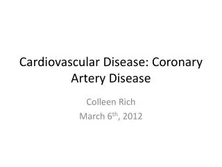
Pathophysiology of Coronary Artery Disease. Blood supply to the heart. Coronary Blood Flow : Constant Demand Arteries & veins are located on the surface of the heart, lying within groves called sulci Blood flows through coronaries during the RELAXATION phase (diastole) Why?.

E N D
Blood supply to the heart • Coronary Blood Flow: Constant Demand • Arteries & veins are located on the surface of the heart, lying within groves called sulci • Blood flows through coronaries during the RELAXATION phase (diastole) Why?
Primary Superficial Arteries • Right Coronary • Right Marginal Branch • Left Coronary • Left Anterior Descending • Left Circumflex
Areas of Coronary Artery Perfusion: • LAD: Majority of the left ventricle: Anterior/Inferior • Circumflex (Cx):Inferior and Posterior LV • RCA: RV, Posterior and Septal
Circumflex LCA LAD
Right Marginal Branch
RCA Rt. Marginal
Step 1: Endothelial Injury • Location? coronaries, carotids, renal arteries, lower extremities • Tunica Intima damaged (How?) • LDL Oxidation: Initiates Inflammatory Process: • Monocytes: Attracted and “glued” to endothelium by “ELAMS” (endothelial-leukocyte adhesion molecules)
STEP 2: Inflammation gone Haywire • Monocytes/Leukocytes enter the Sub-endothelial space • Initiate Smooth Muscle Cell proliferation • Attract more LDL to create a “fatty streak”
Ouch! Blood Clot Normal Narrowed Occluded
LDL’s and Atherosclerosis • Oxidized LDLs act as a “free radical”, which induces endothelial injury inflammation begins • Some LDLs are removed by macrophages, but high LDLs will cause much injury • LDLs = accelerated atherosclerosis
Vessel Lumen Smooth Muscle
Myocardial Oxygen Supply • Cardiac cells extract most of the oxygen delivered (high A-vO2 diff) • Thus additional oxygen can only be met by delivering more blood by the coronary arteries • A reduction in coronary artery lumen size attenuates blood flow
Degree of Occlusion & Blood Flow Blood Flow 25 50 75 100%
Blood Flow • Big reduction beyond 75% occlusion • Severity of disease depends on # of occluded arteries • Also location of occlusion (left coronary vs apical)
Myocardial O2 demand (MVO2) depends on.. • Myocardial tension (pressure x volume) • Inotropic State (Measure?) • Chronotropic state (Measure?) • Myocardial mass
Indirect measure of MVO2 • Rate pressure product (a.k.a. double product, tension-time index) • Considers 2 of the MVO2 indices: • HR X SBP • Good estimate of oxygen use by the heart. • Used to determine angina threshold
End Results of Atherosclerosis Unstable Angina Stable Angina Acute Myocardial Infarction Sudden Cardiac Death
Stable Angina • Angina occurs at a consistent and predictable level of MVO2 • Reduced Coronary Blood Flow • Always exercise at an intensity below the angina threshold • How would you identify the angina threshold?
Anginal Symptoms • Varied: Chest pressure/heaviness • Back, neck, shoulder ache • Diaphoresis • Pallor • Dyspnea • Does it occur upon exertion?
Unstable Angina • Due in part to intra-coronary spasm • This reduces lumen diameter • May occur ANY time (unpredictable) and at any work intensity • Termed “Vasospastic” or “Prinzmetal’s Angina”
Acute Myocardial Infarction • Intra-lumenal thrombus formation • Thrombus lodges in coronary artery, stops blood flow • Cells downstream are starved of O2 • Leads to tissue necrosis
LAD: Thrombosis Area of Infarct
Sudden Death • May be due to the death of heart tissue from large vessel occlusion • or.. emboli (ischemia) induce ventricular arrhythmias that will kill (i.e. ventricular fibrillation)
Infarction symptoms • Similar to angina: diaphoretic, pallor complexion, pain • Vomiting, dyspnea • Symptoms are often ignored or denied by the patient • Odds of survival are greatest if they get help within 1 hour
Complications of MI • Cardiac Tamponade: Fluid between pericardium/myocardium • Pericarditis: Inflammation of the pericardium • Emboli: From either MI thrombus or atrial clots formed with atrial pooling
Most Common Complications: • Congestive Heart Failure: • 75% of MI’s experience overt CHF Fluid backs up… • 25% of MI’s experience “compensated” CHF reduced perfusion to “vital organs”? • Dysrhythmias:The importance of ECG monitoring post-MI
Assignment: • Read: “Cardiovascular disease and the endothelium” • Answer: How does understanding the mechanism of the disease (pathophysiology) improve prevention, detection and intervention strategies?