Download
1 / 2
20 likes | 39 Vues
Chang Liver cells<br><br>Cells of this line contain HeLa marker chromosomes, and were derived via HeLa contamination. The cells are positive for keratin by immunoperoxidase staining.u00a0<br><br>https://www.creative-bioarray.com/Chang-Liver-CSC-C3553-item-39308.htm<br><br>Chang Liver cells, Chang, Liver cells, Chang, Liver, cells<br><br>Cells of this line contain HeLa marker chromosomes, and were derived via HeLa contamination. The cells are positive for keratin by immunoperoxidase staining.u00a0<br><br>https://www.creative-bioarray.com/Chang-Liver-CSC-C3553-item-39308.htm<br><br>Chang Liver cells, Chang, Liver cells, Chang, Liver, cells

E N D
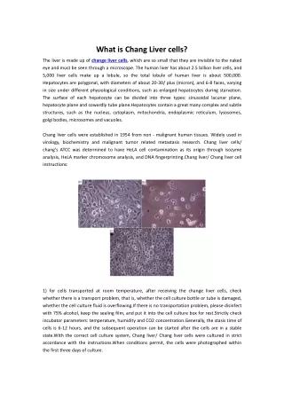
What is Chang Liver cells? The liver is made up of change liver cells, which are so small that they are invisible to the naked eye and must be seen through a microscope. The human liver has about 2.5 billion liver cells, and 5,000 liver cells make up a lobule, so the total lobule of human liver is about 500,000. Hepatocytes are polygonal, with diameters of about 20-30/ plus (micron), and 6-8 faces, varying in size under different physiological conditions, such as enlarged hepatocytes during starvation. The surface of each hepatocyte can be divided into three types: sinusoidal lacunar plane, hepatocyte plane and cowardly tube plane.Hepatocytes contain a great many complex and subtle structures, such as the nucleus, cytoplasm, mitochondria, endoplasmic reticulum, lysosomes, golgi bodies, microsomes and vacuoles. Chang liver cells were established in 1954 from non - malignant human tissues. Widely used in virology, biochemistry and malignant tumor related metastasis research. Chang liver cells/ chang's ATCC was determined to have HeLA cell contamination as its origin through isozyme analysis, HeLA marker chromosome analysis, and DNA fingerprinting.Chang liver/ Chang liver cell instructions: 1) for cells transported at room temperature, after receiving the change liver cells, check whether there is a transport problem, that is, whether the cell culture bottle or tube is damaged, whether the cell culture fluid is overflowing.If there is no transportation problem, please disinfect with 75% alcohol, keep the sealing film, and put it into the cell culture box for rest.Strictly check incubator parameters: temperature, humidity and CO2 concentration.Generally, the stasis time of cells is 6-12 hours, and the subsequent operation can be started after the cells are in a stable state.With the correct cell culture system, Chang liver/ Chang liver cells were cultured in strict accordance with the instructions.When conditions permit, the cells were photographed within the first three days of culture.
2) for the cells transported by dry ice, please put the frozen tube into a 37C tank for quick thawing immediately after removing it, and gently shake the frozen tube to make it melt completely within 1 minute, and pay attention that the water surface should not exceed the edge of the frozen tube cover, otherwise it is prone to contamination. Then it was resuscitated and the culture medium was changed the next day. The selective effects and mechanisms of Archaisms 275 on human gastric cancer cells sgc-7901, human gastric mucous normal cells ges-1 and Chang liver/ Chang liver cells have been studied experimentally, providing theoretical basis for the clinical application of Acidosis. Sgc-7901 cells, gs-1 and Chang liver/ Chang liver cells were treated with ms-275 at different final concentrations, and the growth inhibition effect of the drug on cells was detected by wst-1 method. Annexation and PI double standard flow cytometry was used to detect the selective killing effect of ms-275 on tumor cells sgc-7901 and normal cells.TUNEL staining was used to observe the apotheosis in tumor cell nucleus.The results showed that ms-275 significantly inhibited the growth of gastric cancer cell sgc-7901, and this effect was time-dependent and dose-dependent. The killing effect of ms-275 was selective and showed no apoptosis promoting effect on normal gs-1 cells and chang liver cells human change's hepatocytes. TUNEL staining showed that ms-275 can induce DNA breakage in the nucleus of sgc-7901 cells, which is an important indicator of cell apotheosis. About Creative Bioarray Creative Bioarray is an innovative biotechnology company whose mission focuses on developing unique technologies that provide global scientists with high quality products and satisfactory services to facilitate the investigation of life science researches. We provide a wide range of high quality normal human and animal cells, cell culture medium and reagents, FISH probes, tissue arrays, microorganisms and equipment. In addition, we also offer series of related services including cell services, biosample services and histology services for the researcher to make their project better and faster.