Download
1 / 16
160 likes | 165 Vues
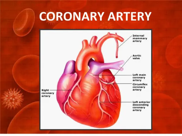
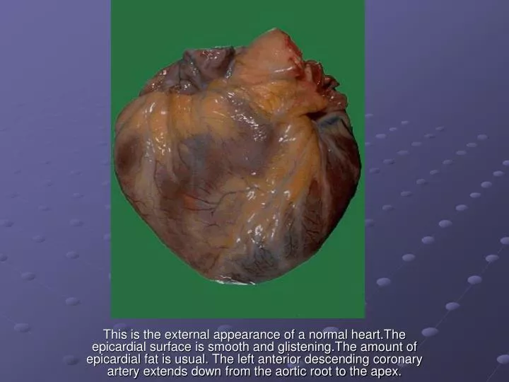
This is the external appearance of a normal heart.The epicardial surface is smooth and glistening.The amount of epicardial fat is usual. The left anterior descending coronary artery extends down from the aortic root to the apex. .

E N D
This is the external appearance of a normal heart.The epicardial surface is smooth and glistening.The amount of epicardial fat is usual. The left anterior descending coronary artery extends down from the aortic root to the apex.
This is the normal appearance of myocardial fibers in longitudinal section. Note the central nuclei and the syncytial arrangement of the fibers, some of which have pale pink intercalated disks.
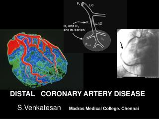
This distal portion of coronary artery shows significant narrowing. Such distal involvement is typical of severe coronary atherosclerosis
This is a high magnification of the aortic atheroma with foam cells and cholesterol clefts.
A coronary artery has been opened longitudinally. The coronary extends from left to right across the middle of the picture and is surrounded by epicardial fat. Increased epicardial fat correlates with increasing total body fat. There is a lot of fat here, suggesting one risk factor for atherosclerosis.
This is the left coronary artery from the aortic root on the left. Extending across the middle of the picture to the right is the anterior descending branch. This coronary shows severe atherosclerosis with extensive calcification. At the far right, there is an area of significant narrowing.
This is coronary atherosclerosis with the complication of hemorrhage into atheromatous plaque, seen here in the center of the photograph. Such hemorrhage acutely may narrow the arterial lumen.
Cross sections of this anterior descending coronary artery demonstrate marked atherosclerosis with narrowing. This is most pronounced at the left in the more proximal portion of this artery. In general, the worst atherosclerosis is proximal,
The anterior surface of the heart demonstrates an opened left anterior descending coronary artery.Within the lumen of the coronary can be seen a dark red recent coronary thrombosis.
At high magnification, the dark red thrombus is apparent in the lumen of the coronary. The yellow tan plaques of atheroma narrow this coronary significantly, and the thrombus occludes it completely.
A thrombosis of a coronary artery is shown here in cross section. This acute thrombosis diminishes blood flow and leads to ischemia and/or infarction, marked clinically by the sudden onset of chest pain.
This myocardial infarction is about 3 to 4 days old. There is an extensive acute inflammatory cell infiltrate and the myocardial fibers are so necrotic that the outlines of them are only barely visible.
This is an intermediate myocardial infarction of 1 to 2 weeks in age. Note that there are remaining normal myocardial fibers at the top. Below these fibers are many macrophages along with numerous capillaries and little collagenization.
The myocardium beneath the endocardial surface at the top demonstrates pale fibrosis with collagenization following healing of a subendocardial myocardial infarction.
An aortic dissection may lead to hemopericardium when blood dissects through the media proximally. Such a massive amount of hemorrhage can lead to cardiac tamponade.
This is infective endocarditis. The aortic valve demonstrates a large, irregular, reddish tan vegetation.Virulent organisms, such as Staphylococcus aureus, produce an "acute" bacterial endocarditis, while some organisms such as Streptococcus viridans produce a "subacute" bacterial endocarditis.