Download
1 / 7
70 likes | 261 Vues
ECG of the Week. This week’s ECG is a challenge? The ECG has findings with significant mortality implications. Are you ready???. Presented by: Bill Rooney, M.D. Obtaining Best Results from this presentation. For best results—please do the following:

E N D
ECG of the Week This week’s ECG is a challenge? The ECG has findings with significant mortality implications. Are you ready??? Presented by: Bill Rooney, M.D.
Obtaining Best Results from this presentation For best results—please do the following: Select “Slide Show” from the menu option on top Select “From the beginning” Slowly click through the presentation Have fun!---Good luck
The request A 51 year old male applies for $1,000,000 of life insurance. There is a history of some generalized fatigue, palpitations, and one recent episode of syncope occurring after exercise which was initially felt to be aggravated by dehydration. One older brother died suddenly a few years back but no specific cause was identified. There has been an echo done recently but the results are not available at the moment. Luckily you do have an ECG to interpret (are you surprised? I think not! This is, of course, an ECG of the week series!!!)
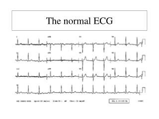
The ECG Here is the ECG!! How would you interpret this tracing?
The interpretation ANSWER: NSR with rate ~72 bpm PR/QRS/QT intervals appear WNL. PR ~ 0.16 sec QRS ~ 0.08 sec QTc ~ 0.42 sec Axis: LAD ~-30o Hypertrophy: None Q waves: Lead III, ? Small r waves in aVF ST-T wave changes: ST segment depression noted in leads I, aVL as well as V4-6 with inverted T waves in I, aVL, V2-6 The echo report is obtained and documents the diagnosis of hypertrophic cardiomyopathy.
Hypertrophic Cardiomyopathy—ECG findings Frequently there are prominent voltages and repolarization changes However, the ECG changes of HCM are not specific to HCM. An echocardiogram is frequently needed to help establish the diagnosis A normal ECG is uncommon! It is seen in <10% of those with HCM!!! Q waves, especially in the inferior and lateral leads are commonly seen. This reflects depolarization of the hypertrophied septum. Deeply inverted T waves are frequently seen—especially in V2-4. Left axis deviation is common. Bi-atrial enlargement is common Hypertrophic Cardiomyopathy Facts to know
Final Thoughts For further information regarding this condition please refer to Dr. Braun’s Housecalls presentation of 6/26/12. The link to this presentation is: http://www.generaliusalifere.com/Publications/Pages/Housecalls.aspx This concludes this week’s ECG of the week. Contact me if you have questions!