
SlideServe
Menu
Préférences
© SlideServe · v2026

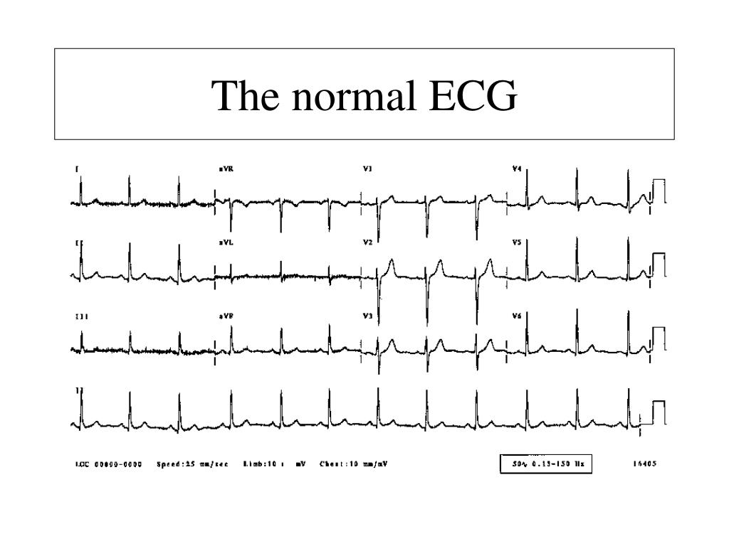
The normal ECG

tahlia avatar
52 diapositives · 2.8K Views · Aug 25, 2014

The normal ECG. Normal sinus rhythm Each p wave followed by a QRS Normal P waves P wave rate 60-100bpm. Sinus bradycardia. Sinus tachycardia. QRS axis. P waves. Height <2.5mm in lead II Width < 11msec in lead II. Right atrial hypertrophy. M shaped p wave in II
The normal ECG - slide 1
01 / 52The normal ECG
1 / 17

Télécharger la présentation

The normal ECG
Une image / un lien ci-dessous est fourni (tel quel) pour télécharger la présentation

Politique de téléchargement : le contenu du site vous est fourni TEL QUEL pour votre information et votre usage personnel ; il ne peut être vendu, concédé sous licence ni partagé sur d’autres sites sans l’accord de son auteur.Le contenu vous est fourni TEL QUEL, pour votre information et votre usage personnel uniquement. Téléchargez la présentation en cliquant sur ce lien. Lors du téléchargement, si vous ne parvenez pas à télécharger une présentation, l’éditeur a peut-être supprimé le fichier de son serveur.Pendant le téléchargement, si vous ne parvenez pas à obtenir une présentation, le fichier a peut-être été supprimé par l’éditeur.

F I N

Plus de contenu associé

Transcription de la présentation
  1. The normal ECG

  2. Normal sinus rhythm • Each p wave followed by a QRS • Normal P waves • P wave rate 60-100bpm

  3. Sinus bradycardia

  4. Sinus tachycardia

  5. QRS axis

  6. P waves • Height <2.5mm in lead II • Width < 11msec in lead II

  7. Right atrial hypertrophy

  8. M shaped p wave in II Prominent negative component in V1

  9. Left atrial hypertrophy M shaped p wave in II Prominent negative component in V1

  10. PR interval • 0.12to 0.2 sec (3-5 small squares)

  11. 2nd degree AV block divided into two types by Mobitz

  12. 2nd degree AV block divided into two types by Mobitz

  13. 2nd degree AV block divided into two types by Mobitz Alias “Wenkebach”

  14. QRS duration • <012sec • If wider then BBB • LBBB (WiLLiaM) • s wave in V1-V3 • rsr pattern in V5-V6 • RBBB (MaRRoW) • rsr in V1

  15. QTc interval (normal 0.42sec) • ST segment • T wave • U wave

  16. Hyperkalaemia

  17. Breathlessness increasing over years ... Difficult

  18. Right ventricular hypertrophy Dominant R in V1 Right axis …… = Mitral stenosis + pulmonary hypertension! + Left atrial abnormality

  19. What now?

  20. Atrial fibrillation with pre-excitation DC Cardioversion or iv procainamide NOT digoxin, verapamil, beta blockers, adenosine...

  21. Inferior myocardial infarction & 4:3 Wenkebach block (i.e. at node)

  22. Trifascicular block Lateral Q’s (A.S.H.) Voltage criteria for LVH HOCM!