Download
1 / 12
140 likes | 1.15k Vues
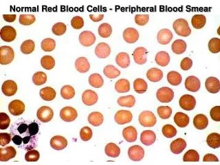
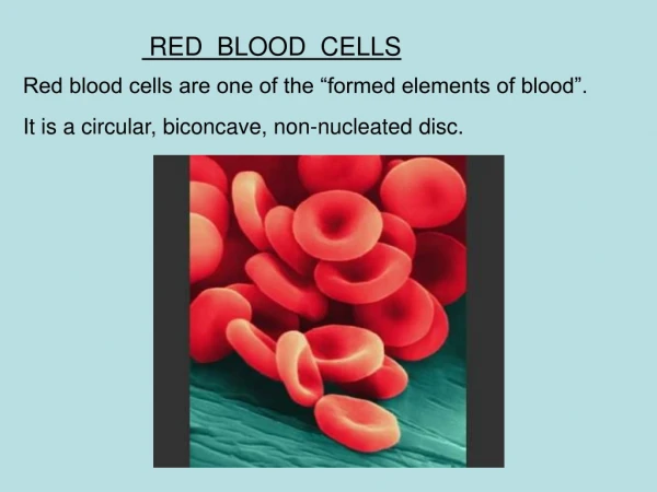
Microscopic Sediment – Red Blood Cells. Red blood cells Pathological finding - cannot appear in filtrate if nephron is intact. result of damage / injury to glomerular membrane, ▸ or urinary tract ▸ strenuous exercise (marathon runners). Microscopic Sediment – Red Blood Cells.

E N D
Microscopic Sediment – Red Blood Cells • Red blood cells • Pathological finding - cannot appear in filtrate if nephron is intact. result of damage / injury to glomerular membrane, ▸ or urinary tract ▸ strenuous exercise (marathon runners)
Microscopic Sediment – Red Blood Cells • differentiate: • Hemoglobinuria – free hemoglobin in urine • Hematuria – presence of intact RBCs in urine • Can you have positive blood on dipstick & negative microscopic for RBCs? • Can you have positive microscopic for RBCs and yet negative dipstick?
Microscopic Sediment – Red Blood Cells • Result of bleeding along urinary tract • Bleeding in nephron – red cell casts formed when the RBCs get caught in precipitating protein • Bleeding in lower GU tract – no protein or casts • 160x mag
Microscopic Sediment – Red Blood Cells • Red Blood Cells • Although NV = 0-2 hpf, an occasional RBC is more significant than occasional WBC. • Detection • High power magnification • Reduced light • yellow - red sheen (sometimes blue-green) • Intact disc or may be crenated • Highly retractile, smooth surface, round • In dilute or alkaline urine appear as ghost or shadow cells
Microscopic Sediment – Red Blood Cells • Urine RBCs can be easily confused with: • Yeast - - generally refract light differently, may have buds, and often are more egg shaped • Bubbles or oil droplets - large variation in size. Even more refractile / and have ‘hard’ appearing edges. • Confirmation – test for hemoglobin - by dipstick, which is most sensitive to free hemoglobin, rather than intact RBCs
Microscopic Sediment – Red Blood Cells • Red Blood Cells • High magnification • Hypertonic urine resulting in some crenated
Microscopic Sediment – Red Blood Cells • RBCs of various shapes & different levels of magnification
Microscopic Sediment – Red Blood Cells • fresh RBCs in the urine are often due to lower urinary tract problems
Microscopic Sediment – Red Blood Cells • When RBCs are subjected to osmotic pressures from having been in the urine for longer periods of time, they become more dismorphic
Microscopic Sediment – Red Blood Cells • RBC can even get small ‘blebs’ on them, making them appear similar to budding yeast.
Microscopic Sediment – Red Blood Cells • Must be differentiated from yeast • @ 160 x