Download
1 / 14
140 likes | 834 Vues
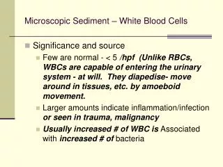
Microscopic Sediment – White Blood Cells. Significance and source Few are normal - < 5 /hpf (Unlike RBCs, WBCs are capable of entering the urinary system - at will. They diapedise- move around in tissues, etc. by amoeboid movement.

E N D
Microscopic Sediment – White Blood Cells • Significance and source • Few are normal - < 5 /hpf (Unlike RBCs, WBCs are capable of entering the urinary system - at will. They diapedise- move around in tissues, etc. by amoeboid movement. • Larger amounts indicate inflammation/infection or seen in trauma, malignancy • Usually increased # of WBC is Associated with increased # of bacteria
Microscopic Sediment – White Blood Cells • Condition of increased WBC in the urine called pyuria / leukocyturia • usually are neutrophils can also be eosinophils or mononuclear - lymphs, monocytes, macrophages, etc. • Addressed on page 79 • May be found in clumps - significant finding, should note on report.
Microscopic Sediment – White Blood Cells • Detection • High power • Fine adjustment • Description • Greyish-blue sheen • @ 10-12 microns in diameter • Fine cytoplasmic granulation, rough surface, may have irregular edges. Nuclei may be mono or poly, but often hard to see detail.
Microscopic Sediment – White Blood Cells • rough surface, may have irregular edges. Nuclei may be mono or poly, but often hard to see detail. • WBC can be confused with RBCs that are swollen, or renal epithelial cells.
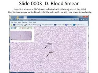
Microscopic Sediment – White Blood Cells • WBC / leukocytes • Higher level of magnification than normally used in routine examination.
Microscopic Sediment – White Blood Cells • WBC • Leukocytes (unstained and Toluidine blue, hpf)
Microscopic Sediment – White Blood Cells • Phase contrast
Microscopic Sediment – White Blood Cells • Pus (degenerated neutrophils) – clumps / aggregates of neutrophils
Microscopic Sediment – White Blood Cells • WBCs and bacteria
Microscopic Sediment – White Blood Cells • Eosinophils • Hansel stain preferred over Wrights to demonstrate presence of eosinophils in urine. • Increases seen in variety of conditions, most notably allergic reactions such as acute graft rejection, schistosomiasis, & acute allergic interstitial nephritis
Microscopic Sediment – White Blood Cells • Lymphocytes • Occasionally seen in normal sediment • Increased numbers reported in acute allergic interstitial nephritis, graft rejection, etc. • Requires special staining to verify identity • Monocytes • Also can be found in above conditions • Also requires special staining to verify identity
Microscopic Sediment – White Blood Cells • Macrophages • Usually of normal size with inclusions in cytoplasm. • Occasionally enlarged with one or more smaller cells engulfed. • Seen in acute inflammatory processes • When filled with fat droplets would be called oval fat bodies.
Microscopic Sediment – White Blood Cells • WBCs, RBCs, cell debris, bacteria