Download
1 / 67
670 likes | 803 Vues
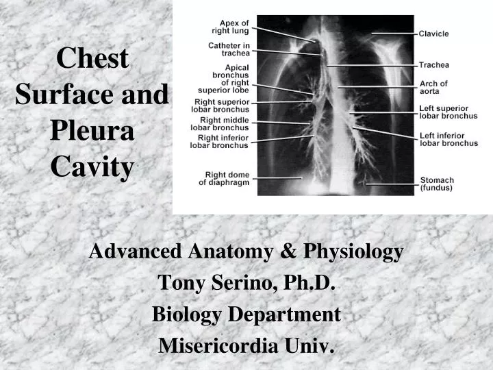
Chest Surface and Pleura Cavity. Advanced Anatomy & Physiology Tony Serino, Ph.D. Biology Department Misericordia Univ. Thoracic Vertebrae. Vertebrae and Ribs. Rib Types and Sternum. Rib Anomalies. Cervical ribs. Bicipital rib (rib fusion). Bifid rib (two heads). 1 st rib. Ribs.

E N D
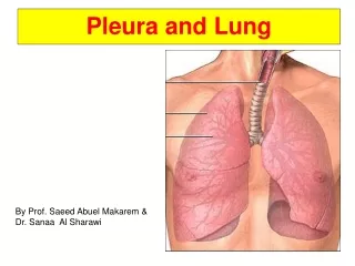
Chest Surface and Pleura Cavity Advanced Anatomy & Physiology Tony Serino, Ph.D. Biology Department Misericordia Univ.
Rib Anomalies Cervical ribs Bicipital rib(rib fusion) Bifid rib (two heads)
1st rib Ribs 2nd rib Crest of head Head Neck Tubercle 11th rib 12th rib
Superficial Muscles Deltopectoral triangle (contains Cephalic vein)
Thoracic Apertures Superior Inferior
Breast Male nipple at T4 Dermatome
Female Breast Retromammary space Suspensory ligaments Tail of breast Glandular tissue and stroma
Female Breast Retromammary space
Blood supply to the Breast Lateral thoracic (from axillary a.) Internal thoracic a.(from subclavian) Anterior intercostals Post. Intercostals(from thoracic aorta) (Venous drainage mostly to axillary v. and internal thoracic v.)
Lymphatic Drainage of Breast Axillary nodes Parasternal nodes Pectoral nodes Subareolar plexus Inferior phrenic nodes
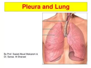
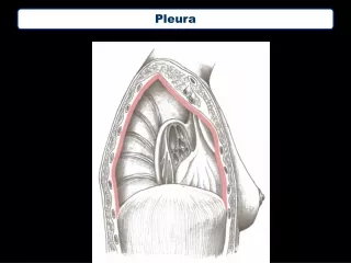
Pleura Cardiac notch Costodiaphragmatic recess Costomediastinal recess
Surface to Deep Structure Alignment Bare Pericardium
Respiration • External Respiration • The exchange of gas between the blood and external environment (usually includes ventilation) • Internal Respiration • The exchange of gas between the blood and the tissues • Cellular Respiration • Burning of fuel to produce energy within cells • Ventilation (Breathing) • Movement of air in and out of the lungs
Respiratory Organs • Divided into: • Upper Respiratory Tract • Includes: nostrils (nares), nasal cavity, and nasopharynx • Lower Respiratory Tract • Includes: larynx, trachea, bronchi, and lungs • Conducting Air passages include: nares to terminal bronchioles • Move air to respiratory membrane • Condition the air • Moisten, Warm, Clean
Bronchi • Primary bronchi lead to to each lung (left and right) • Secondary (lobar) bronchi lead to each lung lobe (3 on right and 2 on left)
Tertiary Bronchi Primary Bronchi Secondary Bronchi Bronchi Branches Tertiary (segmental) bronchi lead to each lung broncho-pulmonary segment Bronchi continue to divide at least 20 more times.
Lung Blood Supply PA PV Note: blood supply to respiratory surface; airway blood supplied bybronchial a. (branch of aorta)
Blood pathways Bronchi PA PV
Bronchioles • Air passages less than 1 mm in diameter are bronchioles. • The terminal bronchioles are the last of the purely conducting air passages.
Alveoli highly specialized for Gas Exchange • Lots of Surface Area • Highly vascular • Thin walls
P = pressure to collapse T = surface tension r = radius Role of surfactant is to decrease surface tension in alveoli.