Download
1 / 41
420 likes | 509 Vues
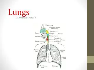
Lungs, pleura. Lungs. Two lungs are soft, spongy and elastic In the child, they are pink, but with age, they become dark and mottled because of the inhalation of dust particles These particles are trapped in the phagocytes of the lung

E N D
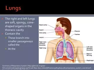
Lungs • Two lungs are soft, spongy and elastic • In the child, they are pink, but with age, they become dark and mottled because of the inhalation of dust particles • These particles are trapped in the phagocytes of the lung • The lungs are situated so that one lies on each side of the mediastinum
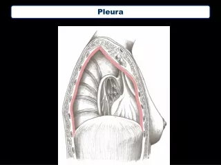
Lungs • Two lungs are separated from each other by the heart and great vessels and other structures in the mediastinum • Each lung is conical, covered with visceral pleura and suspended free in its own pleural cavity • Attached to the mediastinum only by its root
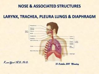
Lungs • Separated from each other by the heart and other structures in the mediastinum • Each lung enclosed by double-layered pleural membrane • Parietal pleura – lines wall of thoracic cavity • Visceral pleura – covers lungs themselves • Pleural cavity is space between layers • Pleural fluid reduces friction, produces surface tension (stick together) • Cardiac notch – heart makes left lung 10% smaller than right
Anatomy of Lungs • Lobes – each lung divides by 1 or 2 fissures • Each lobe receives it own secondary (lobar) bronchus that branch into tertiary (segmental) bronchi • Lobules wrapped in elastic connective tissue and contains a lymphatic vessel, arteriole, venule and branch from terminal bronchiole • Terminal bronchioles branch into respiratory bronchioles which divide into alveolar ducts • About 25 orders of branching
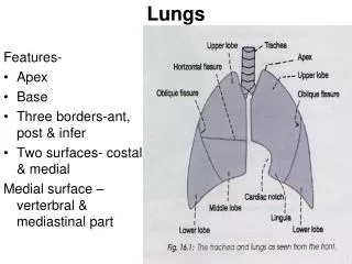
Lungs • A blunt apex projects upward into the neck for about 1 in. (2.5 cm) above the clavicle • A concave base that sits on the diaphragm • A convex costal surface, which corresponds to the concave chest wall • A concave mediastinal surface, which is molded to the pericardium and other mediastinal structures
Lungs • Hilum is a depression in which the bronchi, vessels, and nerves that form the root enter and leave the lung • The anterior border is thin and overlaps the heart • It is here on the left lung that the cardiac notch is found • The posterior border is thick and lies beside the vertebral column
Lobes and Fissures of Rt. Lung • The right lung is slightly larger than the left • Divided by the oblique and horizontal fissures into three lobes: upper, middle, and lower • The oblique fissure runs from the inferior border upward and backward across the medial and costal surfaces until it cuts the posterior border about 2.5 in. (6.25 cm) below the apex
Lobes and Fissures of Rt. Lung • The left lung is divided by a similar oblique fissure into two lobes: upper and lower • There is no horizontal fissure in the left lung
Bronchopulmonary Segment • The bronchopulmonary segments are the anatomic, functional, and surgical units of the lungs • Each lobar (secondary) bronchus gives off branches called segmental (tertiary) bronchi • Each segmental bronchus passes to a structurally and functionally independent unit of a lung lobe called a bronchopulmonary segment, surrounded by connective tissue
Bronchopulmonary Segment • The segmental bronchus is accompanied by a branch of the pulmonary artery • Each segment has its own lymphatic vessels and autonomic nerve supply • On entering a bronchopulmonary segment, each segmental bronchus divides repeatedly
Bronchopulmonary Segment • As the bronchi become smaller, the U-shaped bars of cartilage found in the trachea are gradually replaced by irregular plates of cartilage • The smallest bronchi divide and give rise to bronchioles, which are less than 1 mm in diameter
Bronchopulmonary Segment • Bronchioles possess no cartilage in their walls and are lined with columnar ciliated epithelium • The submucosa possesses a complete layer of circularly arranged smooth muscle fibers
Bronchopulmonary Segment • The bronchioles then divide and give rise to terminal bronchioles which show delicate outpouchings from their walls • Gaseous exchange between blood and air takes place in the walls of these outpouchings, which explains the name respiratory bronchiole
Bronchopulmonary Segment • The diameter of a respiratory bronchiole is about 0.5 mm • The respiratory bronchioles end by branching into alveolar ducts, which lead into tubular passages with numerous thin-walled outpouchings called alveolar sacs
Bronchopulmonary Segment • The alveolar sacs consist of several alveoli opening into a single chamber • Each alveolus is surrounded by a rich network of blood capillaries • Gaseous exchange takes place between the air in the alveolar lumen through the alveolar wall into the blood within the surrounding capillaries
Characteristics of Bronchopulmonary Segment • It is a subdivision of a lung lobe • It is pyramid shaped, with its apex toward the lung root • It is surrounded by connective tissue
Characteristics of Bronchopulmonary Segment • It has a segmental bronchus, a segmental artery, lymph vessels, and autonomic nerves • The segmental vein lies in the connective tissue between adjacent bronchopulmonary segments • Because it is a structural unit, a diseased segment can be removed surgically
Right lung • Superior lobe: Apical, posterior, anterior • Middle lobe: Lateral, medial • Inferior lobe: Superior (apical), medial basal, anterior basal, lateral basal, posterior basal
Left lung • Superior lobe: Apical, posterior, anterior, superior lingular, inferior lingular • Inferior lobe: Superior (apical), medial basal, anterior basal, lateral basal, posterior basal
Blood Supply • The bronchi and the connective tissue of the lung receive their blood supply from the bronchial arteries, which are branches of the descending aorta • The bronchial veins communicate with the pulmonary veins and drain into the azygos and hemiazygos veins
Blood Supply • The alveoli receive deoxygenated blood from the terminal branches of the pulmonary arteries • The oxygenated blood leaving the alveolar capillaries drains into the tributaries of the pulmonary veins • Two pulmonary veins leave each lung root to empty into the left atrium of the heart
Lymph Drainage • The lymph vessels originate in superficial and deep plexuses • They are not present in the alveolar walls • The superficial (subpleural) plexus lies beneath the visceral pleura and drains over the surface of the lung toward the hilum, where the lymph vessels enter the bronchopulmonary nodes
Lymph Drainage • The deep plexus travels to the hilum of the lung and enters the bronchopulmonary nodes • All the lymph from the lung leaves the hilum and drains into the tracheobronchial nodes and then into the bronchomediastinal lymph trunks
Nerve Supply • At the root of each lung is a pulmonary plexus composed of efferent and afferent autonomic nerve fibers • The plexus is formed from branches of the sympathetic trunk and receives parasympathetic fibers from the vagus nerve • The sympathetic efferent fibers produce bronchodilatation and vasoconstriction • The parasympathetic efferent fibers produce bronchoconstriction, vasodilatation, and increased glandular secretion
Alveoli • Cup-shaped outpouching • Alveolar sac – 2 or more alveoli sharing a common opening • 2 types of alveolar epithelial cells • Type I alveolar cells – form nearly continuous lining, more numerous than type II, main site of gas exchange • Type II alveolar cells (septal cells) – free surfaces contain microvilli, secrete alveolar fluid (surfactant reduces tendency to collapse)
Alveolus • Respiratory membrane • Alveolar wall – type I and type II alveolar cells • Epithelial basement membrane • Capillary basement membrane • Capillary endothelium • Very thin – only 0.5 µm thick to allow rapid diffusion of gases • Lungs receive blood from • Pulmonary artery - deoxygenated blood • Bronchial arteries – oxygenated blood to perfuse muscular walls of bronchi and bronchioles
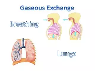
Pulmonary ventilation • Respiration (gas exchange) steps • Pulmonary ventilation/ breathing • Inhalation and exhalation • Exchange of air between atmosphere and alveoli • External (pulmonary) respiration • Exchange of gases between alveoli and blood • Internal (tissue) respiration • Exchange of gases between systemic capillaries and tissue cells • Supplies cellular respiration (makes ATP)
Inhalation/ inspiration • Pressure inside alveoli lust become lower than atmospheric pressure for air to flow into lungs • 760 millimeters of mercury (mmHg) or 1 atmosphere (1 atm) • Achieved by increasing size of lungs • Boyle’s Law – pressure of a gas in a closed container is inversely proportional to the volume of the container • Inhalation – lungs must expand, increasing lung volume, decreasing pressure below atmospheric pressure