Download
1 / 27
310 likes | 711 Vues
Heart and Blood Vessels. By the end of this class you should understand:. The path of blood through the body The basic functions of the blood vessels of the body The purposes of the four chambers of the heart The cardiac cycle and how it can be measured through EKG and pressure.

E N D
By the end of this class you should understand: • The path of blood through the body • The basic functions of the blood vessels of the body • The purposes of the four chambers of the heart • The cardiac cycle and how it can be measured through EKG and pressure
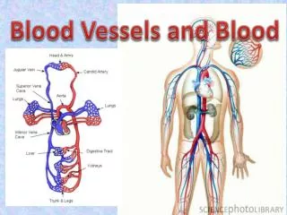
Transportation of Blood • In humans, blood is always contained within blood vessels • If a blood vessel leaks blood into the tissues the result is a bruise • Some types of animals, such as arthropods (insects and spiders) simply bathe their cells in fluid called hemolymph • These blood vessels plus the heart make up the circulatory system
Circulation • All blood is constantly moving along the circulatory system • The most vital nutrient which is used most quickly is oxygen, so blood is usually divided into oxygenated and deoxygenated blood in graphs • Realistically even “deoxygenated” blood usually still has about 25-50% oxygen load
Road Analogy • If cells are houses and must receive service via streets, the largest blood vessels are freeways • Note that like a freeway, major blood vessels such as arteries and veins do NOT deliver oxygen or nutrients but rather move blood quickly to different parts of the body • Almost all nutrient and waste exchange happens in small, leaky blood vessels called capillaries
Arteries and Veins • Arteries move blood away from the heart • Veins move blood towards the heart • Arteries in the body have high pressure and are bright red from the oxygen content • Veins have low pressure and the blood is dark • Deoxygenated blood is NOT blue • It is dark, and vein coverings make it appear blue
Capillaries • All tissues (except cartilage, epidermis and the lens of the eye) have capillaries that blood flows into from the arteries • These capillaries then recollect the blood into veins • Veins are under low pressure so there are valves that keep the blood from flowing backwards • Blood is circulated through veins partially by muscle movement, which is why stretching feels good
Anatomy of Blood Vessels • Major blood vessels (arteries and veins) have smooth muscle and coverings • Smooth muscle helps squeeze blood through and constricts when the blood vessel is damaged • All blood vessels have a smooth inner lining which reduces friction/turbulence • In capillaries these vessels are leaky and so allow nutrients in and out
Variable Flow • Much like actual freeways, where lanes can be opened or closed for more traffic, blood vessels can constrict or dilate to bring more or less blood to parts of the body • Blood flow to skin = “flush” to help cooling • Blood flow to muscles = adrenaline rush • Blood flow to digestive system = food coma
Circulation of Blood • The blood is pumped by the heart through two separate pathways • The left side of the heart is larger than the right because it pumps blood to the entire body • This is why your heart is supposedly on the left side of your chest • The right side pumps blood to the lungs
Heart Chambers • The heart is divided into four chambers, two on the left and two on the right • Entry chamber: atrium (like the entry room to a building) • Main force propeller: ventricle • Division between right and left heart: septum • The heart is two pumps in one, but both contract at the same time
“Lub Dub” Sound • The two sounds of the heart beat are valves closing in the heart to prevent backflow • Heart murmurs are caused by leaky valves • The atria (plural of atrium) contract first to fill the ventricles • The ventricles then start contracting (“lub”) and squeeze blood to the lungs and body, then stop so the atria can refill (“dub”)
Blood Circuits • Blood from the right ventricle goes into the lungs and back to the heart (pulmonary circuit) • Blood from the left ventricle goes to the entire body and back to the heart (systemic circuit) • Systemic circuit is under much higher pressure
Lung Function • The lungs are basically organs full of tiny gas bags wrapped in capillaries • Carbon dioxide is released and oxygen is absorbed by the blood in these capillaries • The blood then goes to the left atrium and ventricle for pumping to the body
Systemic Circuit • Blood leaves the left ventricle through a massive artery called the aorta, which then splits into all the major arteries of the body • Carotid, brachial, renal, iliac, femoral, etc • The arteries then feed into capillaries, which recollect to veins which recover blood to the heart • Largest veins reach the right atrium to begin the pulmonary circuit again
Heart Blood Supply • The heart receives its own blood supply through the coronary arteries • Corona = crown • Arteries can be blocked by excessive body cholesterol • Coronary artery blockage can cut off blood to the heart, causing a heart attack
Cardiac Cycle • The heart goes through a cycle to contract and pump blood through the body efficiently • The cycle is deemed to start when the sinoatrial node (SA node) initiates an action potential • The SA node is the pacemaker of the heart and is influenced by the sympathetic and parasympathetic nervous systems
SA node to AV node • The SA node stimulates the atria to contract, filling the ventricles • The SA node also stimulates the AV node which then stimulates the ventricles • This delay allows the ventricles to fully fill up before initiating ventricular contraction (systole) • If the SA node fails the AV node can take over but the ventricles don’t fill all the way
Ventricular Pressure • When the ventricles contract, they squeeze blood at a high pressure into the major arteries • The pressure in the major arteries is approximately equivalent to the pressure in the heart • 120/80 means peak pressure of 120 when the left ventricle squeezes, 80 when it relaxes • These are also called systole and diastole
Systole and Diastole • Systole is the peak blood pressure caused by the left ventricle squeezing • When the ventricles are refilling it is known as diastole • 120/80 is average healthy blood pressure, higher may indicate unhealthy blood pressure • High blood pressure is known as hypertension and can be caused by diet, lack of exercise, etc
Measuring Blood Pressure • The theory behind measuring blood pressure works on two principles: • The pressure in the major arteries is approximately equivalent to the left ventricle • When the external squeezing pressure on an artery is greater than its blood pressure, the blood flow stops • When the arm cuff cuts off blood pressure entirely, this is systolic pressure • When the arm cuff allows all blood to flow again, this is diastolic pressure • In between these two points, blood only squirts through at diastole but not systole which makes a distinctive sound
Electrocardiogram • The electrocardiogram (ECG/EKG) is the famous “beep beep” in the hospital • Measures the electrical activity of the heart • Small wave corresponds to SA node depolarizing • Large wave corresponds to AV node and ventricles depolarizing • If the SA node fails, only one wave is visible as the AV node takes over and depolarizes the entire heart • Time for an artificial pacemaker!
Enjoy your SPRING BREAK! • Next week is spring break! • Following week lecture is more guts! • Following week lab is the blood lab!