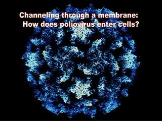
Imaging Poliovirus Entry in Live Cells
130 likes | 296 Vues
This study explores the early stages of poliovirus (PV) infection in live human cells, specifically focusing on how the virus enters the cell and where RNA is released. Using dual-labeled poliovirus and advanced live-cell fluorescence microscopy techniques, including TIRF, we found that RNA release occurs efficiently near the cell membrane after internalization. The release process is ATP-dependent and influenced by temperature, actin, and tyrosine kinases. This work enhances our understanding of PV infection pathways and RNA dynamics in host cells.

Imaging Poliovirus Entry in Live Cells
E N D
Presentation Transcript
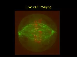
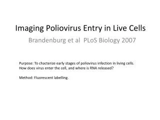
Imaging Poliovirus Entry in Live Cells Purpose: To chacterize early stages of poliovirus infection in living cells. How does virus enter the cell, and where is RNA released? Method: Fluorescent labelling. Brandenburg et al PLoSBiology 2007
Poliovirus (PV) • Simple model system • Infection pathway? • Difficult to characterize virus entry pathways.
Experiment • Dual-labeled PV • RNA: Syto82 (55-65%), green, RNA unfolding reduces signal • Kapsel: Cy5 (100%), red • 300-400 virus particles per cell = 1 pfu/cell • Live-cell fluorescence microscopy • Human cells (HeLa S3)
RNA is released near cell membrane TIRF: Total internal reflection fluorescence microscopy. Isolates nearest 100-200 nm.
Virus is internalizedbefore RNA release pH-sensitive dye: No fluorescence at pH>9 Living cells maintain constant pH => only PV outside cells are pH-sensitive
Neutral red-dependent infectious center (NRIC) assay R78206: blocks RNA release. Brefeldin A: Blocks RNA replication during later stages.
RNA release is ATP dependent NaAz and deoxyglucose depletes cells of ATP
RNA release depends on actin Nocodazole: Inhibits microtubule-dependent transport
Summary • RNA release is rapid and efficient • RNA release takes place after internalization, and near the cell membrane • RNA release depends on temperature, energy (ATP), actin and tyrosine kinases