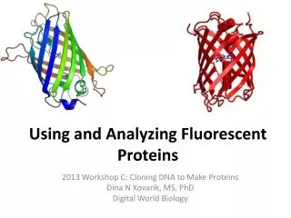
Using and Analyzing Fluorescent Proteins
Using and Analyzing Fluorescent Proteins. 2013 Workshop C: Cloning DNA to Make Proteins Dina N Kovarik, MS, PhD Digital World Biology. Fluorescent Proteins are Valuable Tools. Locate proteins in the cell Track the migration of cells Reporter of expression.

Using and Analyzing Fluorescent Proteins
E N D
Presentation Transcript
Using and Analyzing Fluorescent Proteins 2013 Workshop C: Cloning DNA to Make Proteins Dina N Kovarik, MS, PhD Digital World Biology
Fluorescent Proteins are Valuable Tools • Locate proteins in the cell • Track the migration of cells • Reporter of expression Sister centromeres(green) mark the attachment of microtubules (red) to sister chromatids(blue). Left: Normal. Right: Drug-treated. Mice expressing GFP under UV light (left & right), compared to normal mouse (center). Source: Wikipedia. Source: http://mct.aacrjournals.org/content/2/5.cover-expansion
Mitochondria and Neuronal Injury Fluorescence micrographs (low and high magnification) of a neuron co-transfected with mitochondrially-targeted yellow fluorescent protein and cytoplasmic cyan fluorescent protein. http://www.sfu.ca/rintoul-lab/research.html
In plantaCytometry The proteins are used to mark and consequently identify specific parts of cells - the nuclei and membrane - mapping the development, position and geometry of the cellular make-up in the living plant tissue. http://www.cam.ac.uk/research/news/lighting-up-plant-cells-to-engineer-biology
Using Fluorescent Proteins http://www.scholarpedia.org/article/Fluorescent_proteins
http://www.olympusconfocal.com/applications/fpcolorpalette.htmlhttp://www.olympusconfocal.com/applications/fpcolorpalette.html
Multicolor Labeling of Living HeLa Cell Blue: histone (nucleus). Green: actin (cytoskeleton). Yellow: mitochondria. Orange: golgi. Red: focal adhesions (cell membrane, signal transduction).
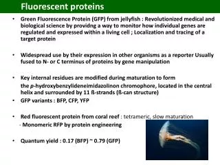
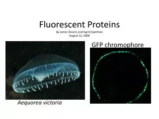
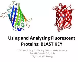
Bioluminescence of the crystal jellyfish, Aequorea victoria 238 amino acid proteins. GFP ribbon diagram. From PDB 1EMA. Source: Wikipedia Source: http://www.conncoll.edu/ccacad/zimmer/GFP-ww/shimomura.html
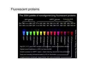
Rainbow of Fluorescent Proteins • “Drawn” with bacteria expressing 8 different fluorescent proteins • Diversity of Mutations Diversity of Colors • “mFruits” • mBlueberry(Blue Fluorescent Protein, or BFP) • mLemon(Yellow Fluorescent Protein, or YFP) • mGrape1 (Cyan Fluorescent Protein, or CFP) • Many others, all with similarly ‘fruity’ names… Source: Wikipedia. http://en.wikipedia.org/wiki/File:FPbeachTsien.jpg
Spectral Diversity of Monomeric FPs Brightness Relative to GFP Emission Wavelength (nm) Chudakov et al, 2010. http://physrev.physiology.org/content/90/3/1103
2Y0G PDB ID 1HUY 2Q57 4AR7 2H5O 2H5Q 3M24 http://www.rcsb.org/pdb/101/motm.do?momID=174
Research Questions The cloning and protein purification experiments you have been conducting in the laboratory involve mTomato, also called red fluorescent protein (RFP). • Is red fluorescent protein (RFP) related to its famous cousin, GFP? • What other fluorescent proteins, if any, are closely related to GFP and/or RFP?