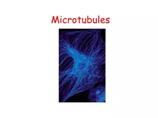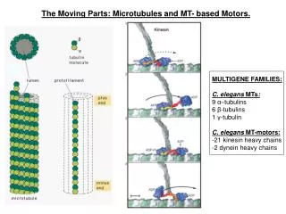
Microtubules (17)
Microtubules (17). Dynamic instability Growing and shrinking microtubules can coexist in the same region of a cell. A given microtubule can switch back and forth between growing and shortening phases. It is an inherent property of the plus end of the microtubule.

Microtubules (17)
E N D
Presentation Transcript
Microtubules (17) • Dynamic instability • Growing and shrinking microtubules can coexist in the same region of a cell. • A given microtubule can switch back and forth between growing and shortening phases. • It is an inherent property of the plus end of the microtubule. • Proteins called +TIPS regulate the rate of growth and shrinkage.
Microtubules (18) • Cilia and Flagella: Structure and Function • Cilia and flagella are hairlike motile organelles. • They have similar structures but different motility. • Cilia tend to occur in large numbers on a cell’s surface.
Microtubules (19) • Cilia and flagella (continued) • Flagella exhibit different beating patterns. • The structure of cilia and flagella contains a central core (axoneme) consisting of microtubules in a 9 + 2 arrangement.
Microtubules (20) • Cilia and flagella (continued) • The basic structure of the axoneme includes a central sheath, connected to the A tubules of peripheral doublets by radial spokes. • The doublets are interconnected to one another by an interdoublet bridge. • A longitudinal view of the axoneme shows the continuous nature of the microtubules
Microtubules (21) • Cilia and flagella (continued) • Cilia and flagella emerge from basal bodies. • The growth of an axoneme occurs at the plus ends of microtubules. • Intraflagellar transport (IFT) is the process responsible for assembling and maintaining flagella. • IFT depends on the activity of both plus end- and minus end-directed microtubules.
Microtubules (22) • The Dynein Arms • The machinery for ciliary and flagellar motion resides in the axoneme. • Ciliary (axonemal) dynein is required for ATP hydrolysis, which supplies energy for locomotion.
Microtubules (23) • The Mechanism of Ciliary and Flagella Locomotion • Swinging cross-bridges generate forces for ciliary or flagellar movement. • Dynein arm of an A tubule binds to a B tubule and undergoes a conformational change that slides tubules past each other. • Sliding alternates from one side of axoneme to another leading to bending.
The Human Perspective: The Role of Cilia in Development and Disease (1) • Situs inversus is a syndrome in which the left-right body symmetry is reversed. • One cause of situs inversus is mutations in the gene encoding ciliary proteins. • Patients with situs inversus suffer from respiratory infections and male infertility.
The Human Perspective: The Role of Cilia in Development and Disease (1) • Many cells have nonmotile primary cilia that sense chemical and mechanical properties of surrounding fluids. • Mutations in primary cilia may lead to polycystic kidney disease. • Cilia are important in developmental processes, and mutations lead to a range of abnormalities.
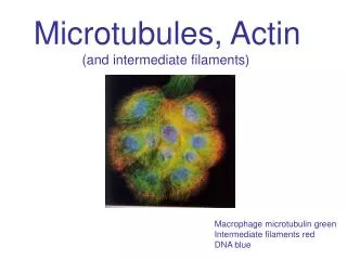
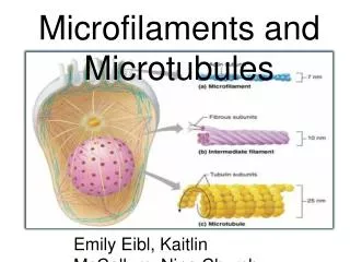
9.4 Intermediate Filaments (1) • Intermediate filaments (IFs)– heterogeneous group of proteins, divided into five major classes. • IFs classes I–IV are used in the construction of filaments; type V (lamins) are present in the inner lining of the nucleus.
Intermediate Filaments (2) • IF Assembly and Disassembly • Assembly: • Basic building block is a rod-like tetramer formed by tow antiparallel dimers. • Both the tetramer and the IF lack polarity. • IFs are less sensitive to chemical agents than other types of cytoskeletal elements.
Intermediate Filaments (3) • Assembly and disassembly of IFs are controlled by phosphorylation and dephosphorylation
Intermediate Filaments (4) • Types and Functions of IFs • IFs containing keratin form the protective barrier of the skin, and epithelial cells of liver and pancreas. • IFs include neurofilaments, which are the major component of the network supporitng neurons.
9.5 Microfilaments (1) • Microfilaments are composed of actin and are involved in cell motility. • Using ATP, actin polymerizes to form actin filaments (“F-actin”). • The two ends of an actin filament have different structural characteristics and dynamic properties.
Microfilaments (2) • One of the micro-filaments appears pointed, and the other appears barbed. • Orientation of the arrowheads formed by actin provides information about direction of the microfilament movement.
Microfilaments (3) • Microfilament Assembly and Disassembly • Actin assembly/disassembly in vitro depends upon concentration of actin monomers. • Filament assembly leads to drop in ATP-actin. • Actin subunits are added to plus end and removed from the minus end (steady state). • Microfilament cytoskeleton is organized by controlling equilibrium between assembly and disassembly of microfilaments.
Microfilaments (4) • Actin polymerization can act as a force-generating mechanism in some cells.
Microfilaments (5) • Myosin: The Molecular Motor of Actin Filaments • All myosins share a characteristic motor head for binding actin and hydrolyzing ATP. • The myosin tail is divergent. • Myosins can be divided into two groups: • Conventional (type II) myosins • Unconventional myosins
Microfilaments (6) • Conventional (Type II) Myosins • They generate force in muscles and some nonmuscle cells. • Each myosin II is composed of two heavy chains, two light chains, and two globular heads (catalytic sites).
Microfilaments (7) • Myosin II (continued) • All of the machinery required for motor activity is contained in a single head. • The tail portion plays a structural role allowing the protein to form filaments.
Microfilaments (8) • Unconventional Myosins • They have only a single head and are unable to assembly into filaments in vitro. • Myosin I’s precise role in cellular activities is unclear. • Myosin V is involved in organelle transport. • Several of them are associated with cytoplasmic vesicles and organelles.