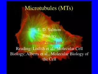
Microtubules
Microtubules. Basic Structure. An -tubulin heterodimer is the basic structural unit of microtubules. Acidic proteins, M.W 55 kDa each Both ά and β tubulin have a nucleotide binding site. -Tubulin has a bound GTP, that does not hydrolyze.
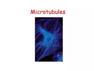

Microtubules
E N D
Presentation Transcript
Basic Structure • An -tubulin heterodimer is the basic structural unit of microtubules. • Acidic proteins, M.W 55 kDa each • Both ά and β tubulin have a nucleotide binding site. • -Tubulin has a bound GTP, that does not hydrolyze. • -Tubulin may have bound GTP or GDP.
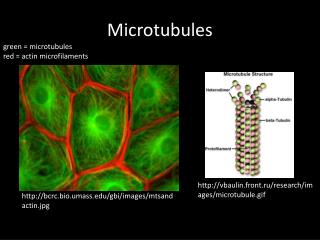
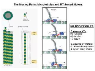
A microtubule is a polymer of ά and ß tubulins. • It is a hollow cylinder, about 24 nm in diameter. • The tubulin heterodimers join end-to-end to form protofilaments, with alternating & subunits. • 13 protofilaments yields a helical arrangement of tubulin heterodimers. Website with an image of a 3-D reconstruction of the structure of an intact microtubule, based on cryo-EM and image processing (by the Visualization Group at Lawrence Berkeley National Laboratory, K. Downing's research group).
These associate laterally to form sheets, & eventually microtubules. • Heterodimers: subunits add at v the plus end, where -tubulin is exposed. • Microtubules can undergo treadmilling, with: • addition of tubulin heterodimers at the plus end • dissociation of tubulin heterodimers at the minus end.
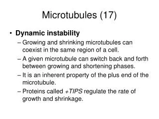
Dynamic instability: • Microtubules may grow steadily & then shrink rapidly by loss of tubulin dimers at the plus end. • A GTP capstabilizes the plus end of a microtubule.
GTP must be bound to both & subunits for a tubulin heterodimer to associate with other heterodimers to form a protofilament. • Subunit addition brings -tubulin that was exposed at the plus end into contact with -tubulin. This promotes hydrolysis of GTP bound to the now interior -tubulin. Pi dissociates. The GTP on -tubulin does not hydrolyze.
Each nucleotide in the protofilament is at an - interface. The inability of GTP to dissociate from the -subunit is consistent with occlusion by a loop from the -subunit. A similar occlusion would account for the inability of -tubulin within a protofilament to exchange GDP/GTP.
View an animation depicting assembly of microtubules. • Then explore the structure of the ,-tubulin heterodimer, using Chime.
Doublet & triplet microtubules: The wall of one microtubule partly consists of the wall of an attached microtubule. • The A tubule is a complete microtubule cylinder, made of 13 protofilaments. • “Piggyback” B or C tubules are made of less than 13 protofilaments, usually 10.
Microtubule Organizing Center (MTOC) • 1. The microtubules in most cells extend outwards from a microtubule organizing center (MTOC) in which the (-) ends of microtubules are anchored. • 2. In animal cells, the major MTOC is the centrosome, located adjacent to the nucleus near the centre of an interphase cell. • 3. The centrosomes of most animal cells contain a pair of centrioles surrounded by amorphous pericentriolar material. • 4. The proteins present: pericentrin, centrin, -Tubulinetc. • 5. -Tubulin, nucleates microtubule assembly within the centrosome.
Role of the MTOC • Control the number of microtubules in a cell • The polarity of the microtubules • The no. of protofilaments that make up the walls of the microtubules. • Time and location of their assembly
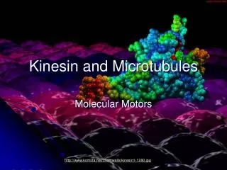
Functions of Microtubules • Centriole structure • Molecular Motors • Cilia and flagella • Mitotic spindle
Centriole Structure • Centrioles: cylindrical structures, found in pairs. • Orientated at right angles to one another. • The wall of each centriole cylinder is made of nine interconnected triplet microtubules.
Centriolar microtubules are relatively stable. • Additional tubulins and other proteins, are either present in centrioles or required for their formation. • They are not found in plants, many unicellular euk. And some animal cells eg. mouse eggs.
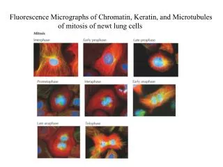
Mitotic Spindle During cell division, the duplicated centrosome helps to organize the mitotic spindle.
During interphase, the centrosome(MTOC) is usually located near the nucleus. Microtubules grow out from the MTOC, forming a hub & spoke array, even during interphase.
With minus endsof most microtubules anchored in the centrosome, microtubules grow & shrink mainly through addition & loss of tubulin heterodimers at their plus ends.
Toxins & Drugs Some toxins and drugs (all of which inhibit mitosis) affect polymerization or depolymerization of tubulin: • Taxol: extracted from the bark of a rare tree, YEW. • Anti-cancer drug, • Binds to and stabilizes microtubules. • Colchicine: extracted from the autmn crocus. • Binds tubulin & blocks polymerization, causes depolymerization.
Nocodazole causes depolymerization of microtubules. • Vinblastine: Anti-cancer drug • Causes depolymerization and formation of vinblastine-tubulin paracrystals.