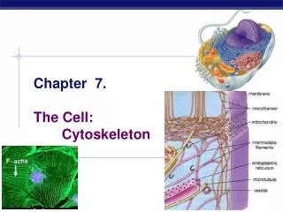
CYTOSKELETON (II) Intermediate filaments and microtubules
510 likes | 2.04k Vues
CYTOSKELETON (II) Intermediate filaments and microtubules. Cell Biology Lecture 10. Readings and Objectives. Reading Russell : Chapter 1 ( incomplete information ) Cooper: Chapter 12 Objectives Intermediate Filaments cytoskeletal scaffolding Microtubules Dynamism

CYTOSKELETON (II) Intermediate filaments and microtubules
E N D
Presentation Transcript
CYTOSKELETON (II)Intermediate filaments and microtubules Cell Biology Lecture 10
Readings and Objectives • Reading • Russell : Chapter 1 (incomplete information) • Cooper: Chapter 12 • Objectives • Intermediate Filaments • cytoskeletal scaffolding • Microtubules • Dynamism • Microtubule Motors and Movement • Organelle trafficking • Cilia and flagella • Mitotic mircotubules
Intermediate Filaments Intermediate filaments (8-11 nm) not directly involved in cell movements provide mechanical strength Supporting scaffold: for organelles and cytoskeleton • Diverse fibrous proteins • Size: 40-200 kD • Not dynamic • Not polar • Regulated by phosphorylation • Six types I-VI
Intermediate Filaments Tissue specific expression Type I and II: keratins, small size (40-70 kD), epithelial cells; Type III: expressed in many cells Vimentin (54 kD), forms a network extending from nucleus to the cell periphery Desmin (53 kD), connects Z-discs in muscle, stabilize actin-myosin Type IV: Neurofilaments; variable size 67, 150 200 kD (Light, Medium, Heavy NF-L, NF-M, NF-H), expressed in mature neurons Type V: nuclear lamins Type VI: Nestins, stem cells, during embryonic development
Structure and Assembly Structure: α-helical rod domain of about 310-350 aa Globular Head and Tail domains, variable size Head and tail domains determine the specific functions
Structure and Assembly Assembly Dimer: central rod form coiled coil Tetramer: staggered antiparallel Protofilament: tetramers assemble end to end Filament: 8 interwoundprotofilament Filaments are stable and nonpolar
Form a network in most cells Extending from a ring surrounding the nucleus to the plasma membrane Associate with cytoskeletal elements Provides a scaffold to organize the internal structure of the cell Cellular arrangement of Intermediate Filaments
Plectin binds actin filaments and microtubules Plectin is 500 kD protein N-terminus domain binds Keratin C-domain binds cytoskeleton bridges them to int. filaments and areas of cell to cell connections Increases the mechanical stability of the cell Interaction with cytoskeleton microtubule Plectin Int. filament anti Plectin A fibroblast cell stained with labelled anti plectin antibody
Known that mechanical stress trigger development of intermediate filaments Transgenic mice expressing mutant Keratin gene (Elain Fuchs lab) severe skinabnormalities Rubbing skincauses blisters Evidence for Cellular Role Elain Fuchs
Human diseases Connection Epidermolysis Bullosa Simplex (EBS) have a keratin gene mutation patients develop skin blisters after minor trauma Amyotrophic lateral Sclerosis (ALS) abnormalities of neurofilaments involving progressive loss of motor neurons muscle atrophy and paralysis
Microtubules are rigid hollow rods (25 nm) Dynamic structures, undergo continual assembly and disassembly Function: cell movements and determining cell shape, organelle transport, mitosis Tubulin, globular protein is the monomer α-tubulin and β-tubulin dimers make up microtubules γ-tubulin in the centrosome plays a critical role in initiating microtubule assembly Microtubules
Microtubules Assembly Tubulin dimers polymerize to form microtubules consisting of 13 protofilaments assembled around a hollow core Protofilaments composed of head-to-tail arrays of tubulin dimers arranged in parallel two distinct ends: a fast-growing + end and a slow-growing minus end
Microtubules Structure Microtubules can undergo treadmilling Tubulin dimers with GTP bound to β-tubulin associate with the growing end GTP is hydrolyzed , tubulin gets less stable, minus end dimers disassociate
Dynamic instability High [tubulin-GTP], dimers added more rapidly than GTP is hydrolyzed microtubule grows Low [tubulin-GTP], GTP at the plus end is hydrolyzed and dimers are lost
Microtubules and Centrosome Centrosome(microtubule organizing centre, MTOC) centrosomeinitiate microtubule growth γ-tubulin as part of γ-tubulin ring complex in centriols required Centriols, Not necessary Microtubules extend to cell cortex Might be stabilized locally MTOC
Regulatory MAPs Modulation of function and stability Post-translational modification of tubulin, eg phosphorylation interactions with microtubule-associated proteins (MAPs) Capping, Severing, disassembling/assembling, end tracking Example: Neurons MT organized differently Axon: - to + Dendrites: bothdirection
Association with motor proteins Kinesins and Dyneins responsible for powering the movements involving microtubules Kinesin: 2 heavy chains, 2 light chains Dynein: 2–3 heavy chainsa number of light and intermediate chains kinesins move towardthe plus end dyneins move towardthe minus end See experiment movie
Higher order molecular machines Cilia and flagellaare microtubule-based projections of the plasma membrane; cell movement very similar structures Cilia beat in a coordinated back-and-forth motion either moves the cell through fluid or moves fluid over the surface of the cell
Higher order molecular machines axoneme of cilia and flagella consists of microtubules in a “9 + 2” pattern: a central pair surrounded by nine outer doublets. Each doublet has a complete A tubule and a B tubule with 10–11 protofilaments, fused to the A tubule Nexin links the microtubules, two arms of dynein are attached to each A tubule
Higher order molecular machines The minus ends of the microtubules are anchored in a basal body, It contains nine triplets of microtubules. Basal bodies serve to initiate growth of axonemal microtubules and to anchor cilia and flagella to the surface of the cell
Higher order molecular machines Movement of cilia and flagella: sliding of outer microtubule doublets relative to one another, powered by motor activity of the axonemal dyneins Dynein bases bind to A tubules, while the head groups bind to B tubules of adjacent doublets
Mitotic Microtubules Microtubules reorganize during mitosis. Interphase microtubule disassembles free tubulin subunits are reassembled into the mitotic spindle Centrosome duplicates, MTOC forms at opposite poles of the mitotic spindle
Mitotic Microtubules 1. Kinetochore microtubules attach to the condensed chromosomes at the centromeres 2. Chromosomal microtubules connect to the ends of the chromosomes via chromokinesin Polar microtubules stabilized by overlapping with each other in the center of the cell. 4. Astral microtubules extend outward
Mitotic Microtubules Anaphase A—chromosomesmove toward spindle poles Anaphase B kinesins cross-link polar microtubules and move them toward the plus end cytoplasmic dynein moves along astral microtubules in the minus-end direction Poles kept apart