Download
1 / 53
530 likes | 704 Vues
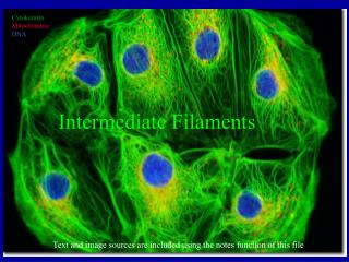
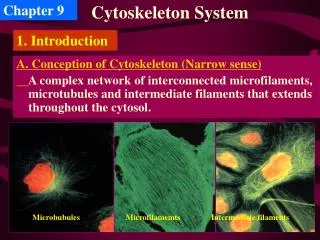
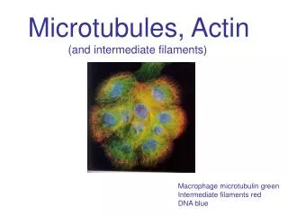
Microtubules, Actin (and intermediate filaments). Macrophage microtubulin green Intermediate filaments red DNA blue. Mitochondria, actin. Cytoplasmic microtubules Cytoplasmic m tubules brkdwn Mitotic spindle Differentiated cell types have stable m tubule structures

E N D
Microtubules, Actin (and intermediate filaments) Macrophage microtubulin green Intermediate filaments red DNA blue
Cytoplasmic microtubules Cytoplasmic mtubules brkdwn Mitotic spindle Differentiated cell types have stable mtubule structures Cilia, flagella form from basal bodies
So what are Microtubules?? … hollow, rigid tubes composed of a/b heterodimers POLAR.. w/o polarity, it can not function.
Each subunit binds GTP … but the beta subunit has intrinsic GTPase activity.
Microtubule networks assemble and disassemble Assembly is initiated at MTOCs MicroTubule Organizing Centers (aka centrosomes)
MTOC 100’s of g subunits serve to nucleate mtubules, Centriole pair (sometimes) Similar to Basal bodies
Growth vs. shrinking is related to the amt. of free tubulin (Cc) Growth when balanced tipped toward >[Cc]
Microtubule assembly and disassemble is CATASTROPHIC!! Occurs preferentially at the plus ends Critical Concentration, Cc, above grows/below shrinks Does not require GTP hydrolysis for assembly The rapid nature of disassembly is due to its constrained internal structure (GDP tubulin)
Autumn Crocus Toolbox: Colchicine (from crocus) binds free tubulin Taxol (from yew) stabilizes microtubules Both cancer therapy drugs, both block mitosis. How? If opposite modes of action? Yew
Microtubules in the mitotic spindle of a mouse fibroblast in culture. Image by Steve Rogers, UNC Dept. Biology. (Confocal Microscope)
Organelles, vesicles with cargo move around the cell. Why’s that?
How do we know? Pulse-chase with radioactive amino acids injected into rat ganglion Assay, slice up axon over time, SDS PAGE autoradiography
Microtubules are needed for trafficking vesicles (10 cm/day) but this is dramatically faster than diffusion in the axon. …in a differentiated cell- dynamic instability is suppressed… by capping proteins.
In vitro, Extruded Exoplasm ATP-dependent
Video microscopy increases resolution and visualizes movement. This movement requires ATP. But AMP-PNP (analog) Competitively inhibits
Ron Vale, UCSF … and AMP-PNP Michael Sheetz, Columbia 17.5 kinesin.mov Movie from Jeff Gelles lab
Video microscopy can be used to track the movement of a single kinesin molecule
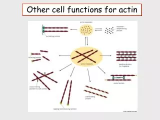
Filaments (F actin) are another important means by which the cytoskeleton is used for trafficking. It is important for cell shape, location and contraction
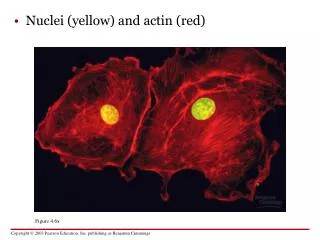
Abundant: 5% of total protein, half as filaments Actin monomers in 4 colors
F actin grows at BOTH ends but the plus ends grows faster. ATP hydrolysis is not required for assembly.
Phalloidin binds F actin, from Death Angel Cytochalasins and lantrunculin bind G actin
Actin filaments are rarely single … Nets and bundles. Profilin thymosin filamin gelsolin myosin
Actin-related proteins promote branching More distally
Racs (Cdc42 is a member) are small GTPases related to the Ras members discussed before. These can be activated by recptors and in turn activate actin assembly
Myosin is a motor that runs along (or tugs at) actin. Actin-based movement of vesicles Myosin-driven cell shape changes Muscle contraction Myosin I and myosin I
Dimer 300 heads here, bind actin
Best understood example … contraction in muscle cells. Multinucleate (formed by cell fusions 50 mm diameter and centimeters long)
Each filament has ~300 heads. Each head binds 5X/s 32 mm in 0.1 s Attached, No ATP, “rigor” Released, binds ATP Cocked, hydrolysis Loose Pi Power stroke, loose ADP Attached again,