Download
1 / 15
150 likes | 295 Vues
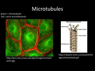
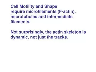
Cell Motility and Shape require microfilaments (F-actin), microtubules and intermediate filaments. Not surprisingly, the actin skeleton is dynamic, not just the tracks. Fibroblasts migrate through tissues by extending forward, attaching to the substrate at focal adhesions and

E N D
Cell Motility and Shape require microfilaments (F-actin), microtubules and intermediate filaments. Not surprisingly, the actin skeleton is dynamic, not just the tracks.
Fibroblasts migrate through tissues by extending forward, attaching to the substrate at focal adhesions and pulling along the tail. Actin assembly and disassembly is the mechanism. Actin is highly conserved.
G actin assembles into F actin. ATP holds together the two lobes. Polymerization can be done in vitro. ATP is hydrolyzed but this is not required for assembly. F actin depolymerizes back to G actin.
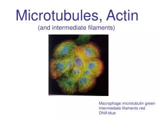
F actin has structural and functional polarity. Can’t be seen directly. Myosin (S1 heads) bind F actin assymetrically.
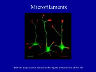
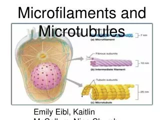
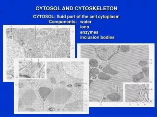
The actin cytoskeleton is organized into bundles and networks to provide framework. Cortical actin is netlike and cytosolic networks are meshes. Cortical nets are attached to the membrane
Actin crosslinkers: Fascin in bundles, Filamin in networks There are many other actin crosslinkers, each with actin-binding domains.
Hypothetical Junctional complex Know components
Left: Platelets at rest Middle: Induced to spread Right: Stellate pattern during clot retraction All are actin based and coupled via ABPs to the actin cytoskeleton
Actin bundles support projections Microvilli (epithelial) and filopodia (fibroblasts)
-3 steps Nucl. Is marked by lag phase Elong by addition at both ends SS at the critical concentration ATP is eventually hydrolyzed so that internal actin is ADP form Growth is greater at + end
Polar growth is demonstrated by decorated nuclei End growth differences due to differences in Cc Cc is measured by capping one end and measuring C for growth G actin ADP, Cc for both ends are equal Treadmilling Tools: cytochalasin D depolymerizes by bind plus ends (fungal alkaloid) Lantrunculin binds G actin and blks polymerization (sponges) Phalloidin, binds F actin and blks Depolmerization (Death Angel)
Summary Cell dynamics are controlled by dynamics in the actin cytoskeleton network Cell shape changes by filament size and treadmilling But shape changes can also occur by actin severing proteins